Review
. 2021 Sep 27;12(10):1519.
doi: 10.3390/genes12101519.

Drosophila Models for Charcot-Marie-Tooth Neuropathy Related to Aminoacyl-tRNA Synthetases

Affiliations
Review

Drosophila Models for Charcot-Marie-Tooth Neuropathy Related to Aminoacyl-tRNA Synthetases

Laura Morant et al. Genes (Basel). .

Abstract

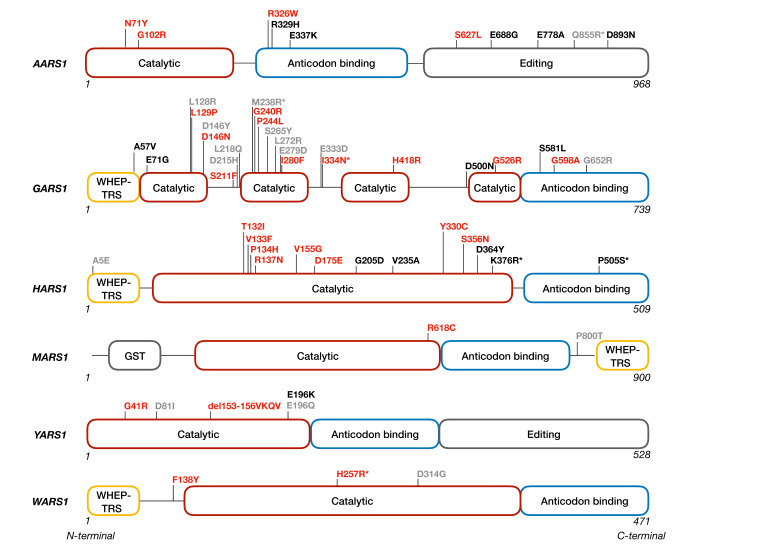
Aminoacyl-tRNA synthetases (aaRS) represent the largest cluster of proteins implicated in Charcot-Marie-Tooth neuropathy (CMT), the most common neuromuscular disorder. Dominant mutations in six aaRS cause different axonal CMT subtypes with common clinical characteristics, including progressive distal muscle weakness and wasting, impaired sensory modalities, gait problems and skeletal deformities. These clinical manifestations are caused by "dying back" axonal degeneration of the longest peripheral sensory and motor neurons. Surprisingly, loss of aminoacylation activity is not a prerequisite for CMT to occur, suggesting a gain-of-function disease mechanism. Here, we present the Drosophila melanogaster disease models that have been developed to understand the molecular pathway(s) underlying GARS1- and YARS1-associated CMT etiology. Expression of dominant CMT mutations in these aaRSs induced comparable neurodegenerative phenotypes, both in larvae and adult animals. Interestingly, recent data suggests that shared molecular pathways, such as dysregulation of global protein synthesis, might play a role in disease pathology. In addition, it has been demonstrated that the important function of nuclear YARS1 in transcriptional regulation and the binding properties of mutant GARS1 are also conserved and can be studied in D. melanogaster in the context of CMT. Taken together, the fly has emerged as a faithful companion model for cellular and molecular studies of aaRS-CMT that also enables in vivo investigation of candidate CMT drugs.

Keywords: Charcot–Marie–Tooth neuropathy; Drosophila melanogaster; aminoacyl-tRNA synthetases; disease-modeling.


Conflict of interest statement

The authors declare no conflict of interest.

Figures

Figure 1
Figure 1
Schematic representation of the location of all CMT-causing dominant mutations reported to date in the domain structure of the corresponding aminoacyl-tRNA synthetases. The CMT mutations can be found in different functional domains. CMT-related mutations affecting the aminoacylation activity are indicated in red, mutations not impairing this activity are depicted in black. The mutations for which the aminoacylation activity remains to be investigated are indicated in grey. The mutations for which the amino acid is not conserved in D. melanogaster are labeled with an asterisk (*). The WHEP-TRS domain refers to a highly conserved helix-turn-helix domain of 46 amino-acids found in some of the aaRSs in higher eukaryotes. GST—Glutathione-S-transferase domain.
Figure 2
Figure 2
Schematic representation of assays used to evaluate aaRSCMT phenotypes in D. melanogaster. Upper panels: assays performed in adult flies. Lower panels: larval assays. The regular and symmetrical organization of the compound eye (red) allows detecting even subtle defects altering the retinal geometry, such as missing bristles and/or merged or misshapen ommatidia (both indicated by red asterisks), resulting in a so-called “rough eye” phenotype [70,71]. The locomotor performance in ageing flies can be evaluated using a negative geotaxis assay (NGA). In this robust behavioral test, the flies are shaken to the bottom of the tube and the climbing speed of the fastest fly to the finish line at 82 mm is measured. The climbing speed is severely decreased in aaRSCMT D melanogaster models. The developmental lethality assay assesses the toxicity of mutant proteins at the whole-organism level by counting the number of offspring expressing the transgene that reaches the adult stage and comparing it to the theoretically expected values. Toxic aaRSCMT mutations induce developmental lethality (red skull) ending the life cycle between the first larval stage and the late pupal stage. The central nervous system of D. melanogaster is composed of the brain and the ventral nerve cord in which the giant fiber interneuron (GF-orange) contains one of the longest axons. It is part of the giant fiber neuronal circuit, mediating a startle response consisting of jump followed by flight. The morphology of the GF can be visualized by filling with fluorescent dye. The GF innervates the tergotrochanteral jump muscle (TTM—yellow) via the TTM motor neuron (blue) and the dorsal longitudinal flight muscle (DLM—brown) via the DLM motor neuron (purple). The giant fiber in aaRSCMT models is characterized by gross morphological defects, such as abnormally thin axonal terminal (red asterisk) with occasional vesicles or constrictions. GFS electrophysiological dysfunctions in aaRSCMT models include reduction in synaptic strength and reliability, characterized by longer response latency (L) and/or smaller amplitude (A) (red arrows) of the output signal after a single stimulation (S) and a decrease in the ability to follow repetitive stimulations at 100 Hz (red asterisks), respectively. The larval neuromuscular junction (NMJ) morphology is a popular model to evaluate the muscle innervation by motor neurons. Each abdominal segment (grey) in the larval body is composed of unique and identifiable muscles (pink) because of their shape, size, and insertion at the larval cuticle. The NMJ morphological analysis consists of visualizing the motor neurons and their connection to the muscle by using pre-(green) and post-(purple) synaptic markers. The phenotype is assessed by measuring the NMJ length (green) and counting the number of synaptic boutons (purple) and branch segments (green). Class IV multidendritic neurons are sensory neurons with the most complex dendritic arborization, capable of sensing multiple noxious stimuli in D. melanogaster larvae. Schematic drawing of class IV multidendritic neurons morphology representing the reduction in dendritic arborization (turquoise) and the decrease in dendritic coverage in the larval body wall due to aaRSCMT mutations.

Similar articles

Cited by

References

    1. Catala M., Kubis N. Gross anatomy and development of the peripheral nervous system. Handb. Clin. Neurol. 2013;115:29–41. doi: 10.1016/B978-0-444-52902-2.00003-5. - DOI - PubMed
    1. Tooth H.H. The Peroneal Type of Progressive Muscular Atrophy. University of Cambridge; London, UK: 1886. - PubMed
    1. Charcot J.M. Sur une forme paticuliere d’atrophie musuculaire progressive souvent familial, debutante par les pieds et les jambes et atteignant plus tard les mains. Rev. Med. Fr. 1886;6:97–138.
    1. Skre H. Genetic and clinical aspects of Charcot-Marie-Tooth’s disease. Clin. Genet. 1974;6:98–118. doi: 10.1111/j.1399-0004.1974.tb00638.x. - DOI - PubMed
    1. Barreto L.C., Oliveira F.S., Nunes P.S., de Franca Costa I.M., Garcez C.A., Goes G.M., Neves E.L., de Souza Siqueira Quintans J., de Souza Araujo A.A. Epidemiologic Study of Charcot-Marie-Tooth Disease: A Systematic Review. Neuroepidemiology. 2016;46:157–165. doi: 10.1159/000443706. - DOI - PubMed

Publication types

MeSH terms

Substances

LinkOut - more resources