Review
. 2021 Sep 29;12(10):1553.
doi: 10.3390/genes12101553.

Insulin-like Growth Factor 1 Signaling in Mammalian Hearing

Affiliations
Review

Insulin-like Growth Factor 1 Signaling in Mammalian Hearing

Ángela García-Mato et al. Genes (Basel). .

Abstract

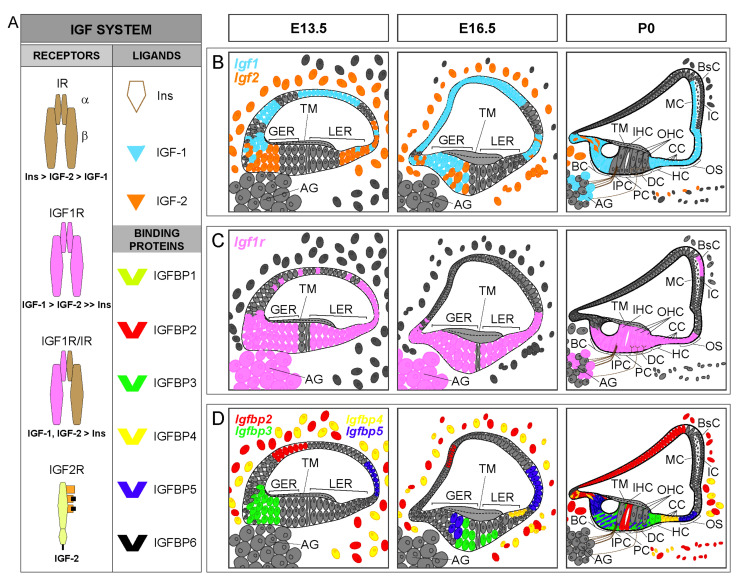
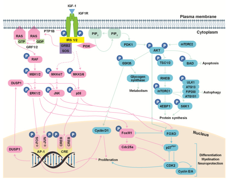
Insulin-like growth factor 1 (IGF-1) is a peptide hormone belonging to the insulin family of proteins. Almost all of the biological effects of IGF-1 are mediated through binding to its high-affinity tyrosine kinase receptor (IGF1R), a transmembrane receptor belonging to the insulin receptor family. Factors, receptors and IGF-binding proteins form the IGF system, which has multiple roles in mammalian development, adult tissue homeostasis, and aging. Consequently, mutations in genes of the IGF system, including downstream intracellular targets, underlie multiple common pathologies and are associated with multiple rare human diseases. Here we review the contribution of the IGF system to our understanding of the molecular and genetic basis of human hearing loss by describing, (i) the expression patterns of the IGF system in the mammalian inner ear; (ii) downstream signaling of IGF-1 in the hearing organ; (iii) mouse mutations in the IGF system, including upstream regulators and downstream targets of IGF-1 that inform cochlear pathophysiology; and (iv) human mutations in these genes causing hearing loss.

Keywords: AKT; Ageing; GH; IGF system; IGF1 mutations; RAF; hearing loss; inner ear; neurodegeneration; rare diseases.


Conflict of interest statement

The authors declare no conflict of interest.

Figures

Figure 1
Figure 1
IGF system expression patterns in the developing cochlea. (A). Schematic representation of the IGF system and receptors binding affinity. The IGF system consists of three ligands (insulin, IGF-1 and IGF-2), transmembrane receptors [insulin receptor (IR), IGF receptors type 1 (IGF1R) and 2 (IGF2R) and hybrid receptor (IGF1R/IR)], and six high affinity IGF-binding proteins (IGFBPs 1–6). (BD) Cartoons of the mouse organ of Corti at embryonic day E13.5, E16.5 and postnatal day P0, showing available cochlear IGF system expression patterns: Igf1 (blue), Igf2 (orange), Igf1r (pink), Igfbp2 (red), Igfbp3 (green), Igfbp4 (yellow) and Igfbp5 (purple). In (D), the expression of Igfbp3 in supporting cells overlaps with the expression of Igfbp5 at P0 (green cells with purple lines). GER, greater epithelial ridge; LER, lesser epithelial ridge; IHC, inner hair cells; OHC, outer hair cells; BC, border cells; IPC, inner phalangeal cells; PC, pillar cells; DC, Deiters’ cells; HC, Hensen’s cells; CC, Claudius’ cells; OS, outer sulcus cells; AG, auditory ganglion; TM, tectorial membrane; MC, marginal cells; IC, intermediate cells; BsC, basal cells. Data have been compiled from: (A) [1,2,3] and (BD) [8,9,10,11,12,13,16].
Figure 2
Figure 2
IGF-1 signaling. Activation of IGF1R can result in signaling via three main pathways: PI3K/AKT, ERK/MAPK and stress kinases (p38 and JNK). The PI3K/AKT pathway modulates cell survival, autophagy, protein synthesis and glucose metabolism; the ERK/MAPK pathway regulates cell growth and differentiation, among other processes; and the stress kinase (p38 and JNK) cascades regulate stress response, proliferation, differentiation, migration and survival. Kinase actions are strictly regulated by phosphatases. PTP1B, protein tyrosine phosphatase 1B; IRS, insulin receptor substrate; GRB2, growth factor receptor-bound protein 2; SOS, son of sevenless; RAS, rat sarcoma; RAF, rapidly accelerated fibrosarcoma; ERK, extracellular signal-regulated kinase; MAPK, mitogen-activated protein kinase; JNK, c-JUN N-terminal kinases; PDK1, phosphoinositide-dependent protein kinase 1; PIP2, phosphatidylinositol 4,5-bisphosphate; PIP3, phosphatidylinositol (3,4,5)-trisphosphate; PI3K, phosphatidylinositol 3-kinase; AKT, thymoma viral proto-oncogene; mTORC1, mammalian target of rapamycin complex 1; mTORC2, mammalian target of rapamycin complex 2; GSK3β, glycogen synthase kinase-3β; TSC1, tuberous sclerosis complex-1; TSC2, tuberous sclerosis complex-2; RHEB, RAS homolog enriched in brain; ULK1, unc-51-like kinase 1; ATG13, mammalian autophagy-related gene 13; FIP200, focal adhesion kinase family-interacting protein of 200 kDa; ATG101, mammalian autophagy-related gene 101; S6K1, p70S6 Kinase 1; 4EBP, eukaryotic translation initiation factor 4E-binding protein; FOXO, forkhead box O; FOXM1, forkhead box protein M1. Data have been compiled from [18,19,23,27,29,30,35,36,37,38].

References

    1. LeRoith D., Holly J.M.P., Forbes B.E. Insulin-like Growth Factors: Ligands, Binding Proteins, and Receptors. Mol. Metab. 2021:101245. doi: 10.1016/j.molmet.2021.101245. - DOI - PMC - PubMed
    1. Bach L.A. IGF-Binding Proteins. J. Mol. Endocrinol. 2018;61:T11–T28. doi: 10.1530/JME-17-0254. - DOI - PubMed
    1. Allard J.B., Duan C. IGF-Binding Proteins: Why Do They Exist and Why Are There So Many? Front. Endocrinol. 2018;9:117. doi: 10.3389/fendo.2018.00117. - DOI - PMC - PubMed
    1. Noguchi G.M., Huising M.O. Integrating the Inputs That Shape Pancreatic Islet Hormone Release. Nat. Metab. 2019;1:1189–1201. doi: 10.1038/s42255-019-0148-2. - DOI - PMC - PubMed
    1. Müller E.E., Locatelli V., Cocchi D. Neuroendocrine Control of Growth Hormone Secretion. Physiol. Rev. 1999;79:511–607. doi: 10.1152/physrev.1999.79.2.511. - DOI - PubMed

Publication types

Substances