. 2021 Sep 27;14(10):983.
doi: 10.3390/ph14100983.

Endothelial Thioredoxin-Interacting Protein Depletion Reduces Hemorrhagic Transformation in Hyperglycemic Mice after Embolic Stroke and Thrombolytic Therapy

Affiliations

Endothelial Thioredoxin-Interacting Protein Depletion Reduces Hemorrhagic Transformation in Hyperglycemic Mice after Embolic Stroke and Thrombolytic Therapy

Mohd Salman et al. Pharmaceuticals (Basel). .

Abstract

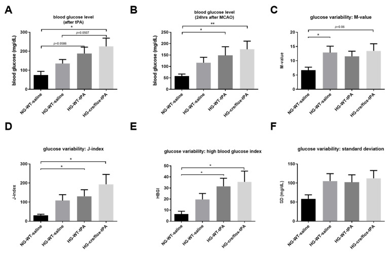
We hypothesize that endothelial-specific thioredoxin-interacting protein knock-out (EC-TXNIP KO) mice will be more resistant to the neurovascular damage (hemorrhagic-transformation-HT) associated with hyperglycemia (HG) in embolic stroke. Adult-male EC-TXNIP KO and wild-type (WT) littermate mice were injected with-streptozotocin (40 mg/kg, i.p.) for five consecutive days to induce diabetes. Four-weeks after confirming HG, mice were subjected to embolic middle cerebral artery occlusion (eMCAO) followed by tissue plasminogen activator (tPA)-reperfusion (10 mg/kg at 3 h post-eMCAO). After the neurological assessment, animals were sacrificed at 24 h for neurovascular stroke outcomes. There were no differences in cerebrovascular anatomy between the strains. Infarct size, edema, and HT as indicated by hemoglobin (Hb)-the content was significantly higher in HG-WT mice, with or without tPA-reperfusion, compared to normoglycemic WT mice. Hyperglycemic EC-TXNIP KO mice treated with tPA tended to show lower Hb-content, edema, infarct area, and less hemorrhagic score compared to WT hyperglycemic mice. EC-TXNIP KO mice showed decreased expression of inflammatory mediators, apoptosis-associated proteins, and nitrotyrosine levels. Further, vascular endothelial growth factor-A and matrix-metalloproteinases (MMP-9/MMP-3), which degrade junction proteins and increase blood-brain-barrier permeability, were decreased in EC-TXNIP KO mice. Together, these findings suggest that vascular-TXNIP could be a novel therapeutic target for neurovascular damage after stroke.

Keywords: embolic stroke; endothelial-specific TXNIP deletion; hemorrhagic transformation; hyperglycemia; tissue plasminogen activator.


Conflict of interest statement

The authors declare that they have no conflict of interest.

Figures

Figure 1
Figure 1
(A) Experimental timeline, (B) analysis of mortality rate, (C) weight loss and (D) neurobehavioral deficits after embolic stroke. Kruskal–Wallis test plus Dunn’s multiple comparisons; WT: wild-type; NG: normoglycemia; HG: hyperglycemia; tPA: tissue-type plasminogen activator; mean ± SEM; n = 7–10.
Figure 2
Figure 2
Analysis of STZ-based hyperglycemic profiling, and glucose variability. (A) EC-TXNIP KO HG mice have significantly higher glycemic level compared to WT HG after tPA-reperfusion (Kruskal–Wallis test plus Dunn’s multiple comparison, Mann–Whitney U test). (B) The tPA treatment may facilitate the maintenance of STZ-induced sub-acute hyperglycemic condition, and EC-TXNIP inhibition may exert a synergistic effect with tPA treatment in this regard (Kruskal–Wallis test plus Dunn’s multiple comparison). (C) The glucose variability was assessed via M-value. STZ-induced diabetic condition should undergo a higher glycemic variation in the first 24 h after stroke (Kruskal–Wallis test plus Dunn’s multiple comparison, Mann–Whitney U test). (D) The glucose variability was assessed via J-index. STZ-induced hyperglycemic condition should undergo a higher glycemic variation (Kruskal–Wallis test plus Dunn’s multiple comparison). The presence or absence of tPA treatment made no difference in this regard. (E) The glucose variability was assessed via standard deviation. STZ-induced hyperglycemic condition in EC-TXNIP KO mice should undergo a higher glycemic variation (Kruskal–Wallis test plus Dunn’s multiple comparison), whereas tPA treatment seems to be suppressive in this regard. (F) The prognosis of hyperglycemia was evaluated via high blood glucose index. STZ-induced hyperglycemia in EC-TXNIP KO as well as wild type mice treated with tPA is more likely to develop hyperglycemia when compared with non-STZ treated wild-type counterparts (Kruskal–Wallis test plus Dunn’s multiple comparison). WT: wild-type; NG: normoglycemia; HG: hyperglycemia; tPA: tissue-type plasminogen activator; mean ± SEM; * p < 0.05 & ** p < 0.01; n = 7–10.
Figure 3
Figure 3
The impact of STZ-induced sub-acute HG in EC-TXNIP KO mice on edema formation, infarct size and HT after eMCAO and tPA-reperfusion. (A) The TTC-stained brain coronal slices were arrayed and digitized. Aligned images are the most representative for each group. (B) Higher level of brain edema was found in the HG group with tPA reperfusion than that NG and HG. However, EC-TXNIP KO mice showed a lower brain edema compared with the HG group with tPA-reperfusion. (C) As for the infarct size, the STZ-induced sub-acute HG leads to a significantly larger infarction. The deleterious effect due to HG, albeit insignificant, should not be disturbed by the absence of TXNIP in endothelial cells. (D) The qualitative hemorrhage scoring suggests that tPA treatment upon hyperglycemic condition led to more severe HT after stroke, and the absence of EC-TXNIP induces non-significant changes. (E,F) tPA-treatment following HG in EC-TXNIP KO mice exhibits a tendency of reduced ipsilateral hemoglobin content. When it comes to hemoglobin excess, the tendency no longer exists. Kruskal–Wallis test plus Dunn’s multiple comparisons; WT: wild-type; NG: normoglycemia; HG: hyperglycemia; HT: hemorrhagic transformation; tPA: tissue-type plasminogen activator; mean ± SEM; * p < 0.05 & ** p < 0.01; n = 7–9.
Figure 4
Figure 4
The impact of STZ-induced sub-acute HG in EC-TXNIP KO on TJs proteins and IgG expression after eMCAO and tPA-reperfusion. (A) Representative Western blot images of ZO-1, Claudin-5, and Occludin. (B) EC-TXNIP deletion non-significantly increased the expression of junctional proteins in post-stroke condition. (C) The expression level of IgG decreased in EC-TXNIP deletion mice when compared to the HG group mice with tPA reperfusion. The optical density of protein bands was analyzed and normalized to β-Actin (unpaired t-test). WT: wild-type; NG: normoglycemia; HG: hyperglycemia; tPA: tissue-type plasminogen activator; mean ± SD; ns = non-significant; n = 6.
Figure 5
Figure 5
The impact of STZ-induced sub-acute HG in EC-TXNIP KO on MMP-9, MMP-3 and VEGFA expression after eMCAO and tPA-reperfusion. (A) Representative Western blot images of MMP-9, MMP-3 and VEGFA. (B) Bar graphs. EC-TXNIP deletion significantly decreased the expression of MMP-9 compared with HG group with tPA treatment. A non-significant downregulation was observed in MMP-3 and VEGFA proteins. The optical density of protein bands was analyzed and normalized to β-Actin (unpaired t-test). WT: wild-type; NG: normoglycemia; HG: hyperglycemia; tPA: tissue-type plasminogen activator; mean ± SD; ns = non-significant; * p < 0.05; n = 6.
Figure 6
Figure 6
The impact of STZ-induced sub-acute HG in EC-TXNIP KO on inflammatory markers, cleaved-caspase-1 and nitrotyrosine after eMCAO and tPA-reperfusion. (A) Representative Western blot images of NLRP3, TNF-α, IL-1β, cleaved-caspase-1 and (B) bar graph. EC-TXNIP KO mice showed a downregulation in the TNF-α, IL-1β, cleaved-caspase-1proteins compared with the HG group with tPA treatment. While no changes were observed with respect to NLRP-3 protein compared with HG group with tPA treatment. (C) Representative image of immunoblot and bar graph of nitrotyrosine. EC-TXNIP KO mice showed a significantly downregulation nitrotyrosine level compared with HG group with tPA treatment. The optical density of protein bands was analyzed and normalized to β-actin (unpaired t-test). WT: wild-type; NG: normoglycemia; HG: hyperglycemia; tPA: tissue-type plasminogen activator; mean ± SD; ns = non-significant; * p < 0.05; n = 6.
Figure 7
Figure 7
The impact of STZ-induced sub-acute HG in EC-TXNIP KO on cleaved PARP-1 and caspase-3 after eMCAO and tPA-reperfusion. (A) Representative Western blot images of cleaved PARP-1, cleaved-caspase-3 and (B) bar graphs. EC-TXNIP mice showed a significant downregulation of cleaved-caspase-3 level compared to HG plus tPA treatment group. While cleaved PAPR-1 was non significantly down regulated compared to HG group with tPA. The optical density of protein bands was analyzed and normalized to β-Actin (unpaired t test). WT: wild-type; NG: normoglycemia; HG: hyperglycemia; tPA: tissue-type plasminogen activator; mean ± SD; ns = nonsignificant * p < 0.05; n = 6.
Figure 8
Figure 8
Schematic diagram illustrating postulated mechanism by which EC-TXNIP KO provides protection against HG with tPA reperfusion following embolic stroke: A variety of different activated pathways including inflammation, apoptotic cascades and MMPs activation, TJs breakdown, and BBB-damage all belong to the HG, which further trigger followed by tPA-reperfusion. However, EC-TXNIP deletion attenuates neuronal cell death and dysfunction through the inhibition of these pathways following HG with tPA-reperfusion in embolic stroke model in mice. BBB: blood-brain barrier; HG: hyperglycemic; MMPs: matrix metalloproteinases TXNIP: thioredoxin-interacting protein; tPA: tissue plasminogen activator.

References

    1. Gaillard T., Miller E. Guidelines for Stroke Survivors With Diabetes Mellitus. Stroke. 2018;49:e215–e217. doi: 10.1161/STROKEAHA.118.020745. - DOI - PubMed
    1. Zhang G., Kim S., Gu X., Yu S.P., Wei L. DPP-4 Inhibitor Linagliptin is Neuroprotective in Hyperglycemic Mice with Stroke via the AKT/mTOR Pathway and Anti-apoptotic Effects. Neurosci. Bull. 2020;36:407–418. doi: 10.1007/s12264-019-00446-w. - DOI - PMC - PubMed
    1. Capes S.E., Hunt D., Malmberg K., Pathak P., Gerstein H.C. Stress Hyperglycemia and Prognosis of Stroke in Nondiabetic and Diabetic Patients. Stroke. 2001;32:2426–2432. doi: 10.1161/hs1001.096194. - DOI - PubMed
    1. Nedergaard M. Transient focal ischemia in hyperglycemic rats is associated with increased cerebral infarction. Brain Res. 1987;408:79–85. doi: 10.1016/0006-8993(87)90360-X. - DOI - PubMed
    1. Ismael S., Nasoohi S., Yoo A., Ahmed H.A., Ishrat T. Tissue Plasminogen Activator Promotes TXNIP-NLRP3 Inflammasome Activation after Hyperglycemic Stroke in Mice. Mol. Neurobiol. 2020;57:2495–2508. doi: 10.1007/s12035-020-01893-7. - DOI - PMC - PubMed