

Mitochondrial Contributions to Hematopoietic Stem Cell Aging
- PMID: 34681777
- PMCID: PMC8537916
- DOI: 10.3390/ijms222011117
Mitochondrial Contributions to Hematopoietic Stem Cell Aging
Abstract
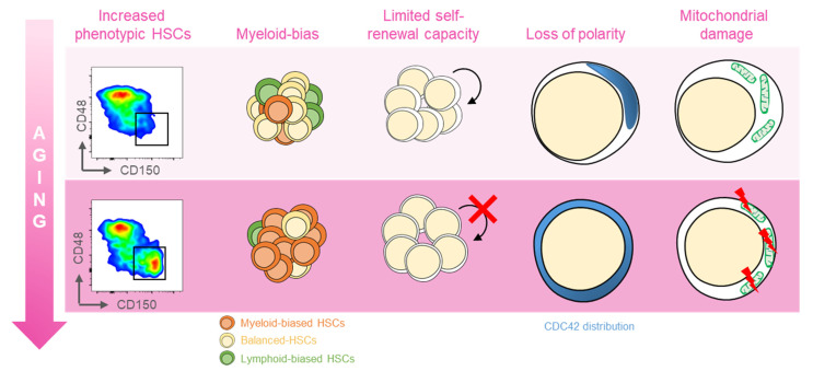
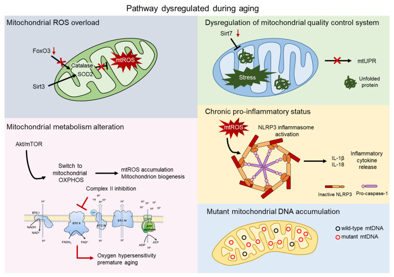
Mitochondrial dysfunction and stem cell exhaustion are two hallmarks of aging. In the hematopoietic system, aging is linked to imbalanced immune response and reduced regenerative capacity in hematopoietic stem cells (HSCs), as well as an increased predisposition to a spectrum of diseases, including myelodysplastic syndrome and acute myeloid leukemia. Myeloid-biased differentiation and loss of polarity are distinct features of aged HSCs, which generally exhibit enhanced mitochondrial oxidative phosphorylation and increased production of reactive oxygen species (ROS), suggesting a direct role for mitochondria in the degenerative process. Here, we provide an overview of current knowledge of the mitochondrial mechanisms that contribute to age-related phenotypes in HSCs. These include mitochondrial ROS production, alteration/activation of mitochondrial metabolism, the quality control pathway of mitochondria, and inflammation. Greater understanding of the key machineries of HSC aging will allow us to identify new therapeutic targets for preventing, delaying, or even reversing aspects of this process.
Keywords: ROS; aging; hematopoiesis; hematopoietic stem cell; inflammation; mitochondrial metabolism; stem cell exhaustion.
Conflict of interest statement
The authors declare no conflict of interest.
Figures
Comment in
-
Metabolic regulation of aged hematopoietic stem cells: key players and mechanisms.Exp Hematol. 2023 Dec;128:2-9. doi: 10.1016/j.exphem.2023.09.006. Epub 2023 Sep 29. Exp Hematol. 2023. PMID: 37778498 No abstract available.
Similar articles
-
Aging of hematopoietic stem cells.Blood. 2018 Feb 1;131(5):479-487. doi: 10.1182/blood-2017-06-746412. Epub 2017 Nov 15. Blood. 2018. PMID: 29141947 Review.
-
Aging of the hematopoietic system.Curr Opin Hematol. 2013 Jul;20(4):355-61. doi: 10.1097/MOH.0b013e3283623c77. Curr Opin Hematol. 2013. PMID: 23739721 Review.
-
Cellular and Molecular Mechanisms Involved in Hematopoietic Stem Cell Aging as a Clinical Prospect.Oxid Med Cell Longev. 2022 Apr 1;2022:2713483. doi: 10.1155/2022/2713483. eCollection 2022. Oxid Med Cell Longev. 2022. PMID: 35401928 Free PMC article. Review.
-
Connexin-43 prevents hematopoietic stem cell senescence through transfer of reactive oxygen species to bone marrow stromal cells.Proc Natl Acad Sci U S A. 2012 Jun 5;109(23):9071-6. doi: 10.1073/pnas.1120358109. Epub 2012 May 18. Proc Natl Acad Sci U S A. 2012. PMID: 22611193 Free PMC article.
-
Cdc42 and aging of hematopoietic stem cells.Curr Opin Hematol. 2013 Jul;20(4):295-300. doi: 10.1097/MOH.0b013e3283615aba. Curr Opin Hematol. 2013. PMID: 23615056 Free PMC article. Review.
Cited by
-
Hematopoietic Stem Cells and the Immune System in Development and Aging.Int J Mol Sci. 2023 Mar 20;24(6):5862. doi: 10.3390/ijms24065862. Int J Mol Sci. 2023. PMID: 36982935 Free PMC article. Review.
-
Pathogenesis and inflammaging in myelodysplastic syndrome.Haematologica. 2025 Feb 1;110(2):283-299. doi: 10.3324/haematol.2023.284944. Haematologica. 2025. PMID: 39445405 Free PMC article. Review.
-
NPM1 ablation induces HSC aging and inflammation to develop myelodysplastic syndrome exacerbated by p53 loss.EMBO Rep. 2022 May 4;23(5):e54262. doi: 10.15252/embr.202154262. Epub 2022 Mar 1. EMBO Rep. 2022. PMID: 35229971 Free PMC article.
-
Modeling GATA2 deficiency in mice: the R396Q mutation disrupts normal hematopoiesis.Leukemia. 2025 Mar;39(3):734-747. doi: 10.1038/s41375-024-02508-z. Epub 2025 Jan 7. Leukemia. 2025. PMID: 39774796 Free PMC article.
-
Role of reactive oxygen species in myelodysplastic syndromes.Cell Mol Biol Lett. 2024 Apr 14;29(1):53. doi: 10.1186/s11658-024-00570-0. Cell Mol Biol Lett. 2024. PMID: 38616283 Free PMC article. Review.
References
Publication types
MeSH terms
Substances
Grants and funding
LinkOut - more resources
Full Text Sources
Medical
