Review
. 2021 Oct 15;22(20):11134.
doi: 10.3390/ijms222011134.

Conserved Structure and Evolution of DPF Domain of PHF10-The Specific Subunit of PBAF Chromatin Remodeling Complex

Affiliations
Review

Conserved Structure and Evolution of DPF Domain of PHF10-The Specific Subunit of PBAF Chromatin Remodeling Complex

Anton O Chugunov et al. Int J Mol Sci. .

Abstract

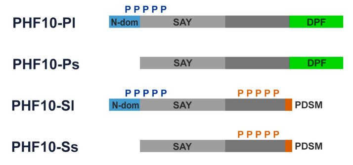
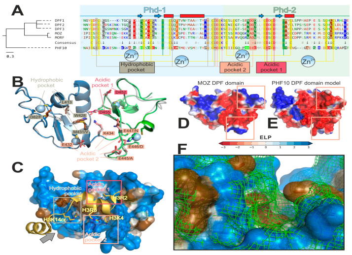
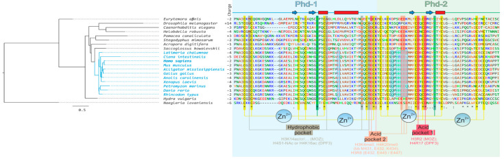
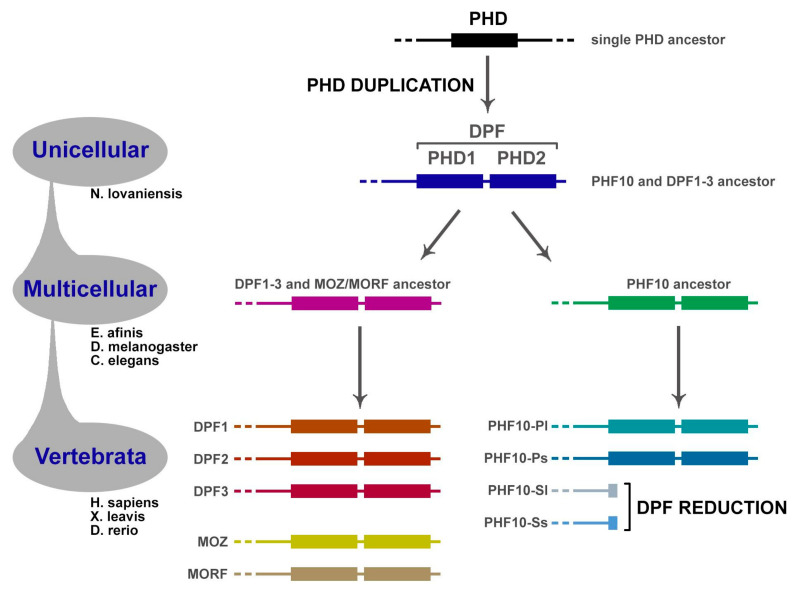
Transcription activation factors and multisubunit coactivator complexes get recruited at specific chromatin sites via protein domains that recognize histone modifications. Single PHDs (plant homeodomains) interact with differentially modified H3 histone tails. Double PHD finger (DPF) domains possess a unique structure different from PHD and are found in six proteins: histone acetyltransferases MOZ and MORF; chromatin remodeling complex BAF (DPF1-3); and chromatin remodeling complex PBAF (PHF10). Among them, PHF10 stands out due to the DPF sequence, structure, and functions. PHF10 is ubiquitously expressed in developing and adult organisms as four isoforms differing in structure (the presence or absence of DPF) and transcription regulation functions. Despite the importance of the DPF domain of PHF10 for transcription activation, its structure remains undetermined. We performed homology modeling of the human PHF10 DPF domain and determined common and distinct features in structure and histone modifications recognition capabilities, which can affect PBAF complex chromatin recruitment. We also traced the evolution of DPF1-3 and PHF10 genes from unicellular to vertebrate organisms. The data reviewed suggest that the DPF domain of PHF10 plays an important role in SWI/SNF-dependent chromatin remodeling during transcription activation.

Keywords: DPF domains; H3K14ac; PBAF; PHD; PHF10; chromatin remodeling; domain evolution; duplication; genes activation.


Conflict of interest statement

The authors declare no conflict of interest.

Figures

Figure 1
Figure 1
PHF10 relative expression in different metacells in a mouse embryo during gastrulation. Meta-cell is a transcriptional state shared by cells from numerous embryos, spanning a specific time range. Scale: log2-fold change (relative to mean expression over all metacells) Data source: [20].
Figure 2
Figure 2
PHF10 isoforms:blue boxes represent 46 amino acids at N-terminus of the long isoforms of PHF10 isoforms; green boxes represent DPF domains; light grey boxes represent SAY domains; orange boxes represent PDSM (phosphorylation-dependent sumoylation motif); blue “P” and orange “P” denote multiple N-terminal phosphorylation and multiple X-cluster phosphorylation, respectively.
Figure 3
Figure 3
PHF10 DPF domain has a number of peculiarities compared to other DPF-domain proteins. (A) Amino acid (a.a.) comparison suggests an early evolutionary divergence of PHF10. Left: phylogenetic tree for six DPF-domain-containing proteins: MOZ and MORF histone acetyltransferases; DPF1–3 and PHF-10 chromatin remodeling complex subunits. Right: corresponding a.a. alignment formatted for better visualization of PHF10 peculiarities. PHD-1 and -2 comprising DPF-domain have light blue and light green background, respectively. Secondary structure (determined for MOZ structure (Pdb ID: 3V43) [38]) is shown above the sequences (blue arrows for β-strands and red rectangles for α-helices). A.a. are colored and grouped according to properties: blue (“+”)—positively charged [Lys, Arg]; red (“−”)—negatively charged [Asp, Glu]; green (“~”)—polar [Ser, Thr, Asn, Gln]; bright-green—His; brown (“#”)—aliphatic [Ala, Val, Leu, Ile, Met]; orange—aromatic non-polar [Phe, Tyr, Trp]; black—Cys, Gly and Pro. Zn2+-binging sites (two per one PHD module) are shown as yellow (Cys) and green (His) vertical stripes. A consensus line is shown between PHF10 and five other sequences. The 100%-conserved positions (with respect to the aforementioned a.a. groups) are pale (except Zn2+-binding sites and histone-binding pockets). The marked dissimilarity in the PHF10 sequence as opposed to the five other proteins is designated by an arrow and a coloured background (depending on the a.a. group) for PHF10 and other proteins (if conserved in all five resting sequences). Three histone-binding pockets are annotated and designated with vertical stripes: 1) hydrophobic (binds non-polar lysine modifications: H3K14ac/cr/bu in MOZ; H4S1-Nac or H4K16ac in DPF3); acidic-1 (100% conserved; anchors H3R2 in MOZ or H4R17 in DPF3); and acidic-2 (binds unmodified H3K4 or H4K20; and anchors H3R8). B–F: Homology model of PHF10 visualizes its peculiarities in 3D (in comparison with MOZ, which was a structural template for MODELLER 9.19). (B) Overview of a model. PHD-1 and -2 are blue and green, respectively. Parent MOZ structure is shown as a semi-transparent gray cartoon. Residues of the four Zn2+-binding sites are represented as sticks and ions as spheres. Residues of the three histone-binding pockets are annotated; corresponding MOZ residue is typed after a slash, if different. Note the dissimilarities in the hydrophobic and acidic-2 pockets, which may determine PHF10 specificity. (C) A visualization of how PHF10 might bind the H3 tail (based on the MOZ/H3K14cr complex (Pdb ID: 5B76) [13]). PHF10 model is shown with a semi-transparent surface colored as a gradient from blue (polar) to brown (non-polar) according to MHP scale [45,46]; the peptide is colored gold. Binding pockets are shown with rectangles: hydrophobic binds non-polar H3K14cr; acidic-1 anchors H3R2 via a salt bridge (red dotted line); acidic-2 recognizes a charged H3K4 via three hydrogen bonds with the backbone of M431, E432, and K434 (shown as pink dotted lines); and anchors H3R8 via salt bridge to E432 and probably E445/E447 (shown as pink red lines). The gray arrow defines a viewpoint for the F panel. (D,E): Comparison of MOZ (D) and PHF10 (E) electrostatic properties. Note that PHF10 has a more negatively charged acidic pocket-2 due to the PHF10-specific HHEEE sequence pattern. (F) Comparison of the hydrophobic pocket’s shape for PHF-10 (colored surface) and MOZ (green mesh). Due to simultaneous substitutions in the pocket’s walls (I382/F and M431/V; after slash is a MOZ residue), it appears to be curved in MOZ (and apparently MORF and DPF1–3), and straighter in PHF10. This may result in an increased preference of PHF10 for bulky hydrophobic modifications at H3K14 or H4K16. Interactive versions of panels B–F of this figure may be downloaded as a PyMol *.pse session from the Supplementary information for this paper.
Figure 4
Figure 4
Amino acid alignment of PHF10 from different species and (probably) several ancestral forms. For details of the figure description, see Figure 3. Chordata in the tree are highlighted in blue. The conserved PHF10 features for this clade are in blue semi-transparent boxes.
Figure 5
Figure 5
Evolution of DPF-containing proteins. Originally, PHF10 and the gene ancestral to DPF13 and MOZ/MORF groups diverged from the common ancestor. Afterwards, the latter group multiplied to five members. Most likely, PHD duplication occurred in the very last eukaryotic common ancestor, since the DPF domain is present in all these genes.

References

    1. Jain K., Fraser C.S., Marunde M.R., Parker M.M., Sagum C., Burg J.M., Hall N., Popova I.K., Rodriguez K.L., Vaidya A., et al. Characterization of the Plant Homeodomain (PHD) Reader Family for Their Histone Tail Interactions. Epigenetics Chromatin. 2020;13:3. doi: 10.1186/s13072-020-0328-z. - DOI - PMC - PubMed
    1. Morrison E.A., Musselman C.A. Chromatin Signaling and Diseases. Academic Press; London, UK: 2016. The Role of PHD Fingers in Chromatin Signaling: Mechanisms and Functional Consequences of the Recognition of Histone and Non-histone Targets; pp. 127–147.
    1. Soshnikova N.V., Sheynov A.A., Tatarskiy E.V., Georgieva S.G. The DPF Domain as a Unique Structural Unit Participating in Transcriptional Activation, Cell Differentiation, and Malignant Transformation. Acta Nat. 2020;12:57–65. doi: 10.32607/actanaturae.11092. - DOI - PMC - PubMed
    1. Yang X.-J. MOZ and MORF Acetyltransferases: Molecular Interaction, Animal Development and Human Disease. Biochim. Biophys. Acta. 2015;1853:1818–1826. doi: 10.1016/j.bbamcr.2015.04.014. - DOI - PubMed
    1. Huang F., Abmayr S.M., Workman J.L. Regulation of KAT6 Acetyltransferases and Their Roles in Cell Cycle Progression, Stem Cell Maintenance, and Human Disease. Mol. Cell. Biol. 2016;36:1900–1907. doi: 10.1128/MCB.00055-16. - DOI - PMC - PubMed

LinkOut - more resources