. 2021 Oct 16;22(20):11179.
doi: 10.3390/ijms222011179.

Platelet-Derived PCSK9 Is Associated with LDL Metabolism and Modulates Atherothrombotic Mechanisms in Coronary Artery Disease

Affiliations

Platelet-Derived PCSK9 Is Associated with LDL Metabolism and Modulates Atherothrombotic Mechanisms in Coronary Artery Disease

Álvaro Petersen-Uribe et al. Int J Mol Sci. .

Abstract

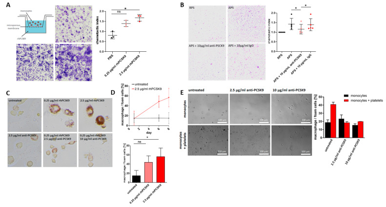
Platelets play a significant role in atherothrombosis. Proprotein convertase subtilisin/kexin type 9 (PCSK9) is critically involved in the regulation of LDL metabolism and interacts with platelet function. The effect of PCSK9 in platelet function is poorly understood. The authors of this article sought to characterize platelets as a major source of PCSK9 and PCSK9's role in atherothrombosis. In a large cohort of patients with coronary artery disease (CAD), platelet count, platelet reactivity, and platelet-derived PCSK9 release were analyzed. The role of platelet PCSK9 on platelet and monocyte function was investigated in vitro. Platelet count and hyper-reactivity correlated with plasma LDL in CAD. The circulating platelets express on their surface and release substantial amounts of PCSK9. Release of PCSK9 augmented platelet-dependent thrombosis, monocyte migration, and differentiation into macrophages/foam cells. Platelets and PCSK9 accumulated in tissue derived from atherosclerotic carotid arteries in areas of macrophages. PCSK9 inhibition reduced platelet activation and platelet-dependent thrombo-inflammation. The authors identified platelets as a source of PCSK9 in CAD, which may have an impact on LDL metabolism. Furthermore, platelet-derived PCSK9 contributes to atherothrombosis, and inhibition of PCSK9 attenuates thrombo-inflammation, which may contribute to the reported beneficial clinical effects.

Keywords: LDL; PCSK9; atherothrombosis; platelets; thrombo-inflammation.


Conflict of interest statement

The authors declare no conflict of interest. The funders had no role in the design of the study; in the collection, analyses, or interpretation of data; in the writing of the manuscript, or in the decision to publish the results.

Figures

Figure 1
Figure 1
Release of PCSK9 from activated platelets. (A) Representative immunoblot images of PCSK9 expression in HepG2 cells, human platelets, and APS (CRP-stimulated, 5 µg/mL) from three independent experiments. (B) Platelet degranulation and release of PCSK9 was verified by surface expression of P-selectin (CD62P) using fluorochrome-conjugated PE-anti-CD62P and FITC-anti-PCSK9 antibodies by flow cytometry. Representative immunohistograms of three independent experiments are shown. PCSK9 signal was not significantly increased after ADP stimulation compared to non-stimulated platelets (n = 3), but CRP stimulation resulted in a statistically significant increase in anti-PCSK9-FITC binding (plotted: mean ± SEM; n = 4, Mann–Whitney test, ns = not significant, * p < 0.05). (C) Correlation of platelet activation and PCSK9 expression. Surface expression of CD62P and of PCSK9 on non-stimulated (upper panel) or CRP-stimulated platelets (1 μg/mL) (lower panel) were determined by flow cytometry. Data indicate Pearson’s correlation coefficient. (D) Effect of LDL on surface expression of PCSK9 on platelets. PRP was incubated with or without 50 µg/mL LDL and thereafter stimulated with CRP (1 µg/mL) or left unstimulated and stained with PCSK9-FITC and analyzed by flow cytometry. Left panel: representative immunofluorescence histograms of three independent experiments are shown. Right panel: LDL treatment had a significant effect on the PCSK9 expression on CRP stimulated platelets (plotted: mean ± SEM, n = 6, Mann–Whitney test, * p < 0.05).
Figure 2
Figure 2
(A) Inhibition of PCSK9 decreased platelet aggregation. PRP was incubated with or without 15 µg/mL anti-PCSK9 (evolocumab). The samples were stimulated with CRP (0.125 µg/mL, 0.25 µg/mL, 0.5 µg/mL) or ADP (1.25 µM, 2.5 µM, 5 µM) and platelet aggregation was measured by light transmission aggregometry. Representative CRP-induced platelet aggregation curves demonstrating the effect of anti-PCSK9 treatment on platelet aggregation. Statistical analysis revealed the percentage of aggregation was reduced by anti-PCSK9 treatment (plotted: mean ± SEM; n ≥ 3, Wilcoxon signed-rank test, ns = not significant, * p < 0.05). (B) Representative ADP-induced platelet aggregation curves demonstrating the effect of anti-PCSK9 treatment on platelet aggregation. Statistical analysis revealed that the percentage of aggregation was reduced by anti-PCSK9 treatment under stimulation with 5 µM ADP (plotted: mean ± SEM; n ≥ 3, Wilcoxon signed-rank test, ns = not significant, * p < 0.05). (C) Anti-PCSK9 reduced platelet-dependent thrombus formation. Human whole blood was perfused over a collagen-coated surface (100 µg/mL) at a shear rate of 1000 s−1. Representative fluorescence images of thrombus formation over time for n ≥ 3 independent experiments. (Scale bar = 100 µm.) (D) Representative end-point fluorescence images for n ≥ 3 independent experiments. (Scale bar = 100 µm.) (E) Statistical analysis of thrombus coverage showed a significant decrease of the thrombus area in whole blood samples treated with anti-PCSK9 antibodies compared to the IgG control and the vehicle control (plotted: mean ± SEM; n ≥ 3, Mann–Whitney test, * p < 0.05). (F) Thrombus formation over 120 s, measured in one representative experiment.
Figure 3
Figure 3
PCSK9 induced platelet migration. (A) Schematic view of experimental set up and representative images for three independent experiments. A significant chemotactic effect of rhPCSK9 (0.25 µg/mL and 2.5 µg/mL) on platelets could be observed (plotted: mean ± SEM; n = 3, Kruskal–Wallis test, * p < 0.05). (B) Representative images of the chemotactic effect of APS compared to resting platelet supernatants (RPS). This effect is prevented by anti-PCSK9 (10 µg/mL) but not by IgG control (plotted: mean ± SEM; n = 5, Mann–Whitney test, * p < 0.05). (C) Platelets propagate monocyte-derived macrophage development via PCSK9. Representative images of macrophage development under stimulation with rhPCSK9 (0.25 µg/mL, 2.5 µg/mL). The effect was inhibited by anti-PCSK9 antibodies (2.5 µg/mL, 10 µg/mL). (D) Macrophage/foam cells development over eight days under rhPCSK treatment (2.5 µg/mL). Statistical analysis of macrophage/foam cells development after eight days treatment with rhPCSK9 (0.25 µg/mL, 2.5 µg/mL) showed significant increase in macrophage/foam cells development (plotted: mean ± SEM; n ≥ 3, biological replicates; Kruskal–Wallis test, * p < 0.05). (E) Representative images of monocytes and monocytes/platelet co-culture. The analysis of macrophage development in monocyte/platelet co-culture after nine days treatment with anti-PCSK9 antibodies (2.5 µg/mL, 10 µg/mL) showed inhibition of macrophage development (plotted: mean ± SEM, n = 2).
Figure 4
Figure 4
(A) CD42b, PCSK9, and CD68 immunostaining of human atherosclerotic carotid tissue. The representative immunostaining photomicrographs of CD42b, PCSK9, and CD68 expression in atherosclerotic tissue derived from endarterectomy specimen from carotid arteries are shown (n = 3). (B) Proposed role of platelet-derived PCSK9 in LDL metabolism and atherothrombosis. Enhanced release of PCSK9 from platelets promotes LDLR degradation and the increase of plasma LDL. Platelet-PCSK9 induces monocyte migration, macrophage development, and enhanced thrombosis.

References

    1. Gawaz M., Langer H., May A.E. Platelets in inflammation and atherogenesis. J. Clin. Investig. 2005;115:3378–3384. doi: 10.1172/JCI27196. - DOI - PMC - PubMed
    1. Gawaz M. Role of platelets in coronary thrombosis and reperfusion of ischemic myocardium. Cardiovasc. Res. 2004;61:498–511. doi: 10.1016/j.cardiores.2003.11.036. - DOI - PubMed
    1. Massberg S., Brand K., Grüner S., Page S., Müller E., Müller I., Bergmeier W., Richter T., Lorenz M., Konrad I., et al. A Critical Role of Platelet Adhesion in the Initiation of Atherosclerotic Lesion Formation. J. Exp. Med. 2002;196:887–896. doi: 10.1084/jem.20012044. - DOI - PMC - PubMed
    1. Geisler T., Schaeffeler E., Dippon J., Winter S., Buse V., Bischofs C., Zuern C., Moerike K., Gawaz M., Schwab M. CYP2C19 and nongenetic factors predict poor responsiveness to clopidogrel loading dose after coronary stent implantation. Pharmacogenomics. 2008;9:1251–1259. doi: 10.2217/14622416.9.9.1251. - DOI - PubMed
    1. DiMinno G., Silver M.J., Cerbone A.M., Rainone A., Postiglione A., Mancini M. Increased fibrinogen binding to platelets from patients with familial hypercholesterolemia. Arter. Off. J. Am. Heart Assoc. Inc. 1986;6:203–211. doi: 10.1161/01.ATV.6.2.203. - DOI - PubMed