

Hodgkin Lymphoma: A Special Microenvironment
- PMID: 34682791
- PMCID: PMC8541076
- DOI: 10.3390/jcm10204665
Hodgkin Lymphoma: A Special Microenvironment
Abstract
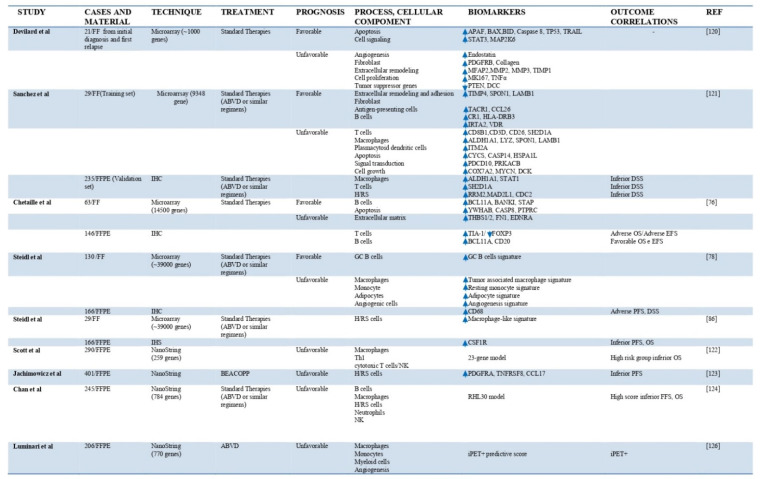
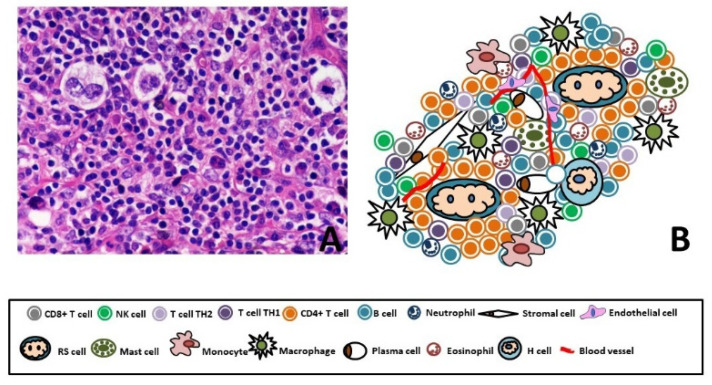
Classical Hodgkin's lymphoma (cHL) is one of the most particular lymphomas for the few tumor cells surrounded by an inflammatory microenvironment. Reed-Sternberg (RS) and Hodgkin (H) cells reprogram and evade antitumor mechanisms of the normal cells present in the microenvironment. The cells of microenvironment are essential for growth and survival of the RS/H cells and are recruited through the effect of cytokines/chemokines. We summarize recent advances in gene expression profiling (GEP) analysis applied to study microenvironment component in cHL. We also describe the main therapies that target not only the neoplastic cells but also the cellular components of the background.
Keywords: Classical Hodgkin’s lymphoma; gene expression profiling; microenvironment; therapy.
Conflict of interest statement
The authors declare no conflict of interest.
Figures
References
-
- Swerdlow S.H., Campo E., Harris N.L., Jaffe E.S., Pileri S.A., Stein H., Thiele J. WHO Classification of Tumours of Haematopoietic and Lymphoid Tissues. Revised 4th ed. Volume 2. International Agency for Research on Cancer; Lyon, France: 2017.
-
- Rengstl B., Newrzela S., Heinrich T., Weiser C., Thalheimer F.B., Schmid F., Warner K., Hartmann S., Schroeder T., Küppers R., et al. Incomplete cytokinesis and re-fusion of small mononucleated Hodgkin cells lead to giant multinucleated Reed-Sternberg cells. Proc. Natl. Acad. Sci. USA. 2013;110:20729–20734. doi: 10.1073/pnas.1312509110. - DOI - PMC - PubMed
-
- Jaffe E.S., Harris N.L., Stein H., Vardiman J. Tumours of Haematopoietic and Lymphoid Tissues. 3rd ed. IARC Press; Lyon, France: 2001.
Publication types
LinkOut - more resources
Full Text Sources
