. 2021 Oct 19;9(10):2172.
doi: 10.3390/microorganisms9102172.

Novel Phage-Derived Depolymerase with Activity against Proteus mirabilis Biofilms

Affiliations

Novel Phage-Derived Depolymerase with Activity against Proteus mirabilis Biofilms

Cormac J Rice et al. Microorganisms. .

Abstract

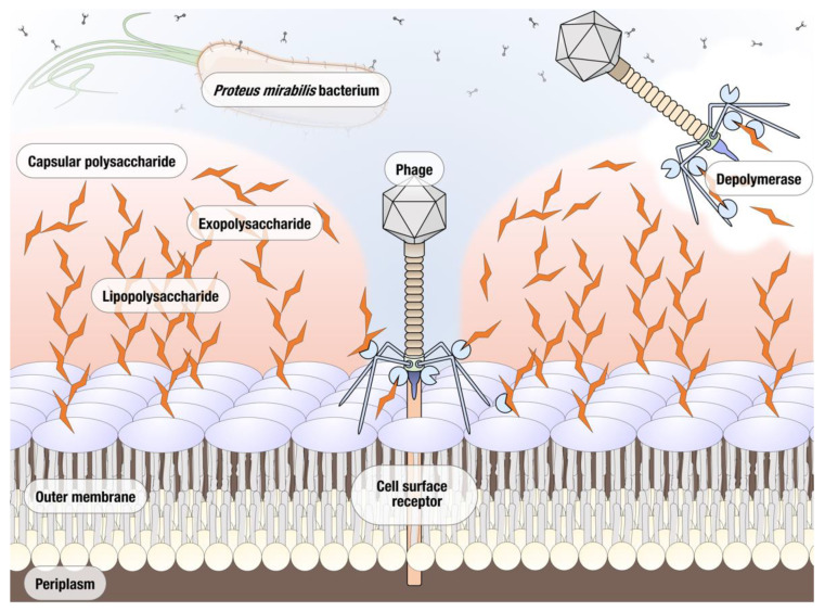
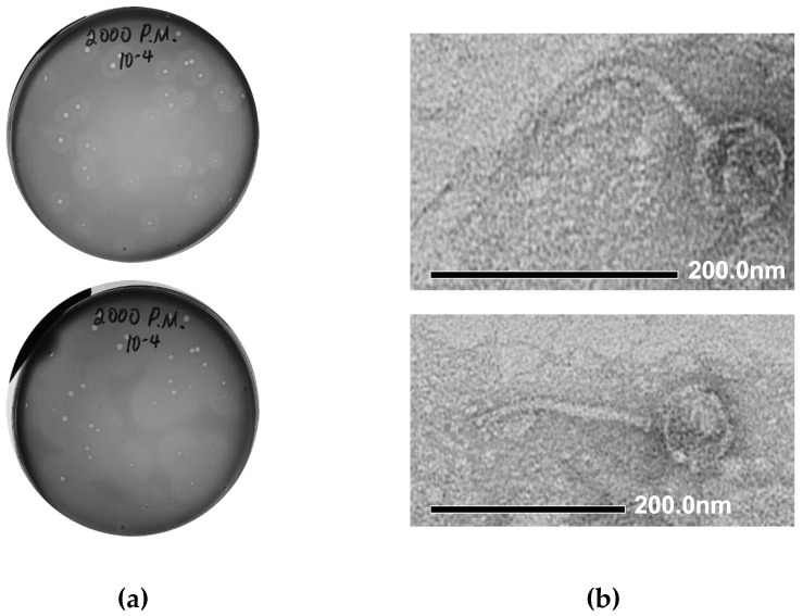
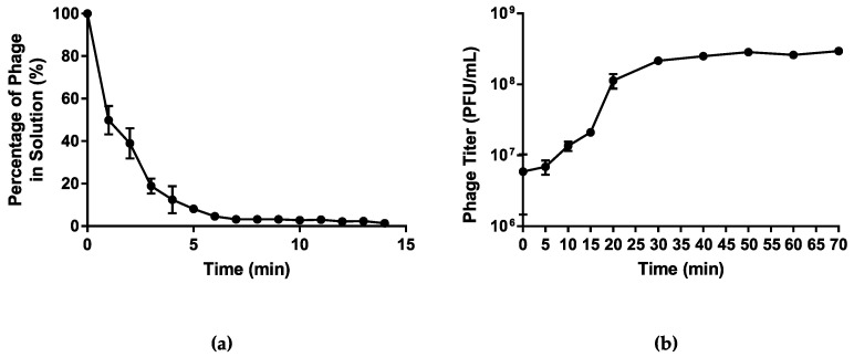
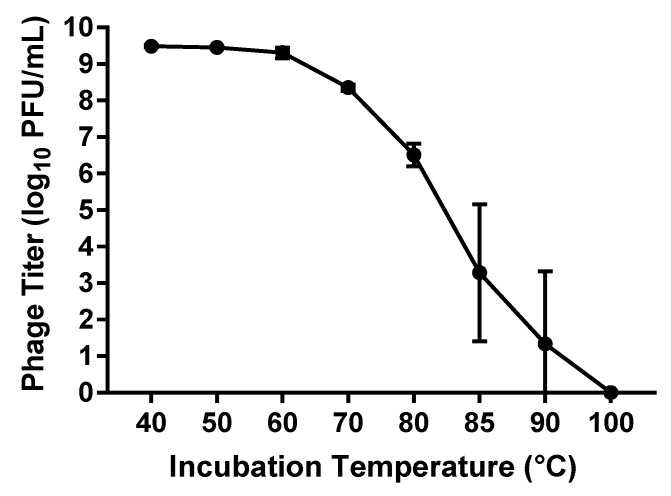
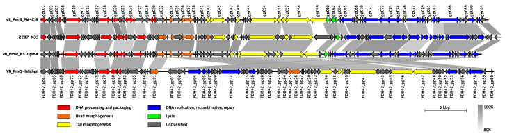
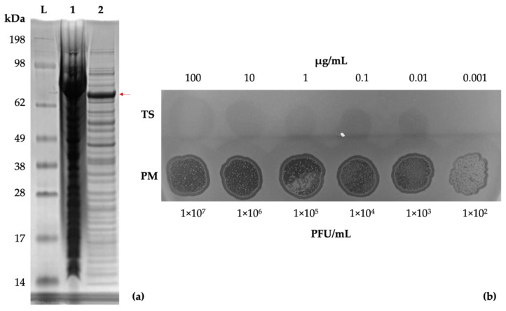
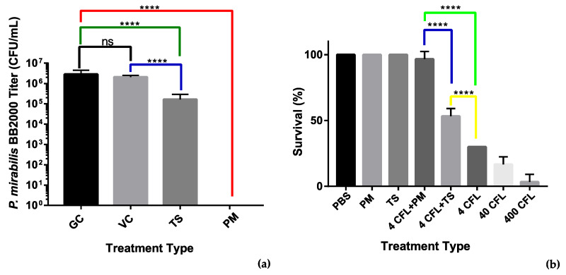
The adherence of Proteus mirabilis to the surface of urinary catheters leads to colonization and eventual blockage of the catheter lumen by unique crystalline biofilms produced by these opportunistic pathogens, making P. mirabilis one of the leading causes of catheter-associated urinary tract infections. The Proteus biofilms reduce efficiency of antibiotic-based treatment, which in turn increases the risk of antibiotic resistance development. Bacteriophages and their enzymes have recently become investigated as alternative treatment options. In this study, a novel Proteus bacteriophage (vB_PmiS_PM-CJR) was isolated from an environmental sample and fully characterized. The phage displayed depolymerase activity and the subsequent genome analysis revealed the presence of a pectate lyase domain in its tail spike protein. The protein was heterologously expressed and purified; the ability of the purified tail spike to degrade Proteus biofilms was tested. We showed that the application of the tail spike protein was able to reduce the adherence of bacterial biofilm to plastic pegs in a MBEC (minimum biofilm eradication concentration) assay and improve the survival of Galleria mellonella larvae infected with Proteus mirabilis. Our study is the first to successfully isolate and characterize a biofilm depolymerase from a Proteus phage, demonstrating the potential of this group of enzymes in treatment of Proteus infections.

Keywords: Proteus; antibiotic resistance; bacteriophage; biofilms; depolymerases; pectate lyase; urinary tract infections (UTIs).


Conflict of interest statement

The authors declare no conflict of interest. The funders had no role in the design of the study; in the collection, analyses, or interpretation of data; in the writing of the manuscript, or in the decision to publish the results.

Figures

Figure 1
Figure 1
Potential polymeric targets for polysaccharide depolymerases of Proteus bacteriophages.
Figure 2
Figure 2
Morphological characteristics of vB_PmiS_PM-CJR. (a) Plaque morphology. The presence of semi-turbid haloes surrounding the plaques proper is visible after 24 h (top). Expansion of haloes after 48 h (bottom). (b) Transmission electron microscopy of phage particles. Phage particles were stained with 1% uranyl acetate and visualized at 40,000× (top) and 30,000× (bottom) magnification.
Figure 3
Figure 3
Growth characteristics of vB_PmiS_PM-CJR. (a) The host adsorption assay. The percentage of the phage that did not adsorb to the host cells is shown on the y axis. (b) One-step growth curve. Phage titer at different times is shown on the y axis, time point 0 corresponds to the moment of the first plating (26 min post-infection). Mean values ± SD (standard deviation) of three independent experiments are plotted.
Figure 4
Figure 4
Thermal stability assay results. The titer (log10 PFU (plaque forming units)/mL) of phage remaining after incubation at temperatures from 40 to 100 °C for 1 h is shown. Mean values ± SD of three independent experiments are plotted.
Figure 5
Figure 5
Genomic organization and whole-genome comparison of vB_PmiS_PM-CJR and closely related phages. Areas of substantial similarity between two genomes are shown as trapezia connecting two genome regions colored according to average nucleotide identity levels of these regions (as calculated by BLASTn). Arrows on the genome map represent identified genes and demonstrate the direction of their transcription. They are colored to reflect common functions of the encoded products. Putative genes with unidentified function are shown in grey color. Original functional annotations were used for each of the genomes.
Figure 6
Figure 6
Tail spike expression and confirmation of activity. (a) SDS-PAGE gel of E. coli KRX cells expressing the recombinant tail spike protein. Lanes: SeeBlue Plus2 reference ladder (L), total protein (1), soluble protein fraction (2). A band of approximately 72 kDa, corresponding to a monomer of the tail spike protein, is visible (indicated with a red arrow). (b) Spot testing (10 µL) of the purified tail spike protein and phage suspension on a lawn of P. mirabilis BB2000.
Figure 7
Figure 7
Analysis of antimicrobial activity of the phage vB_PmiS_PM-CJR and its tail spike. (a) MBEC (minimum biofilm eradication concentration) adherence assay. Treatments: GC—growth control, VC—vehicle control (50 mM Tris-HCl pH 8.0 in LBB), TS—tail spike dialyzed into vehicle, PM—phage vB_PmiS_PM-CJR in 1× PBS. (b) Galleria mellonella infection model. The number of surviving larvae 48 h post-treatment are shown. Treatments (all in 1× PBS): PBS—buffer only, PM—phage only, TS—tail spike only; 4, 40, and 400 CFL—Proteus mirabilis injections of 4, 40, and 400 CFU (colony forming units)/larva. Modifiers +PM and +TS are used to indicate administration of either 2 × 10⁵ vB_PmiS_PM-CJR phage particles or 2 µg of the purified tail spike protein per larva, respectively. Both experiments were conducted in triplicate, mean values ± SD are reported; ns—not significant, ****—p < 0.0001 (one-way ANOVA with Tukey’s post-hoc test).

References

    1. Jacobsen S.M., Stickler D.J., Mobley H.L.T., Shirtliff M.E. Complicated Catheter-Associated Urinary Tract Infections Due to Escherichia coli and Proteus mirabilis. Clin. Microbiol. Rev. 2008;21:26–59. doi: 10.1128/CMR.00019-07. - DOI - PMC - PubMed
    1. Sriwanthana B., Mobley H. Proteus mirabilis urease: Histidine 320 of UreC is essential for urea hydrolysis and nickel ion binding within the native enzyme. Infect. Immun. 1993;61:2570–2577. doi: 10.1128/iai.61.6.2570-2577.1993. - DOI - PMC - PubMed
    1. Milo S., Heylen R.A., Glancy J., Williams G.T., Patenall B.L., Hathaway H.J., Thet N.T., Allinson S.L., Laabei M., Jenkins A.T.A. A small-molecular inhibitor against Proteus mirabilis urease to treat catheter-associated urinary tract infections. Sci. Rep. 2021;11:1–15. doi: 10.1038/s41598-021-83257-2. - DOI - PMC - PubMed
    1. Wasfi R., Hamed S., Amer M.A., Fahmy L.I. Proteus mirabilis Biofilm: Development and Therapeutic Strategies. Front. Cell. Infect. Microbiol. 2020;10:414. doi: 10.3389/fcimb.2020.00414. - DOI - PMC - PubMed
    1. Myelnikov D. An Alternative Cure: The Adoption and Survival of Bacteriophage Therapy in the USSR, 1922–1955. J. Hist. Med. Allied Sci. 2018;73:385–411. doi: 10.1093/jhmas/jry024. - DOI - PMC - PubMed

LinkOut - more resources