Review
. 2021 Sep 23;13(10):1542.
doi: 10.3390/pharmaceutics13101542.

Drug Penetration into the Central Nervous System: Pharmacokinetic Concepts and In Vitro Model Systems

Affiliations
Review

Drug Penetration into the Central Nervous System: Pharmacokinetic Concepts and In Vitro Model Systems

Felix Neumaier et al. Pharmaceutics. .

Abstract

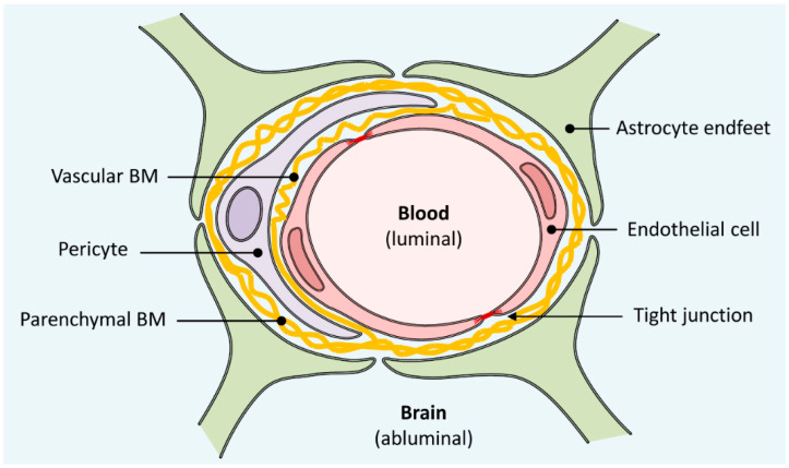
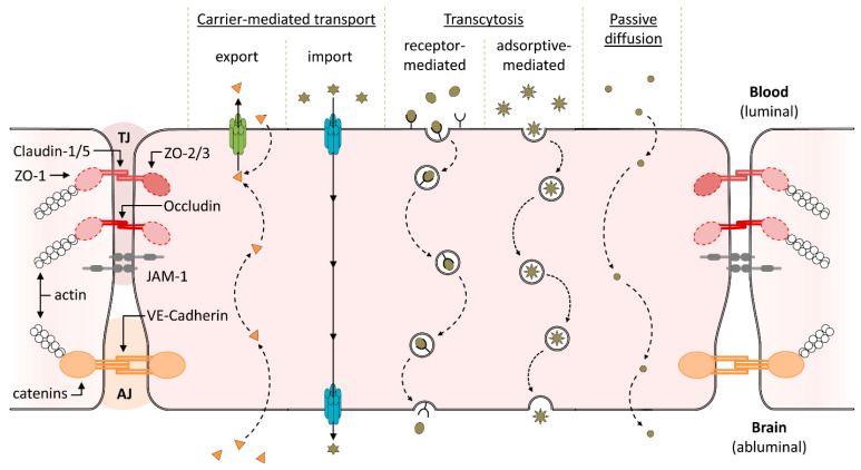
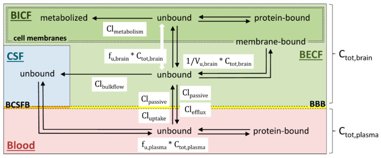
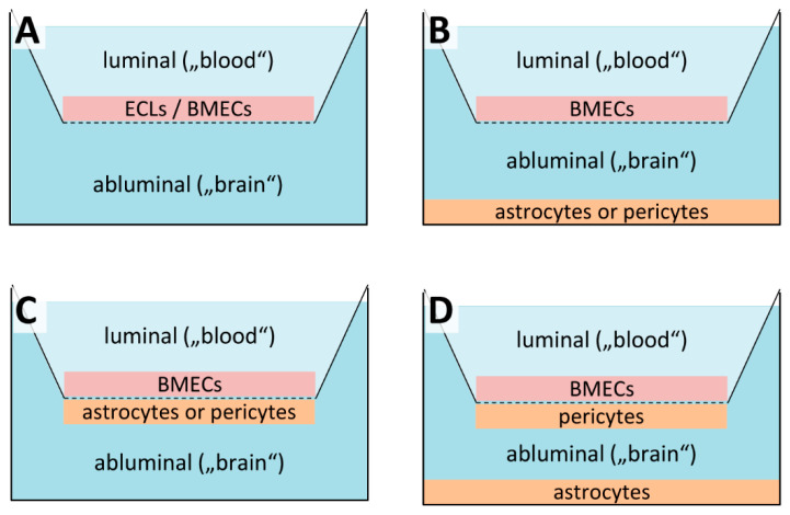
Delivery of most drugs into the central nervous system (CNS) is restricted by the blood-brain barrier (BBB), which remains a significant bottleneck for development of novel CNS-targeted therapeutics or molecular tracers for neuroimaging. Consistent failure to reliably predict drug efficiency based on single measures for the rate or extent of brain penetration has led to the emergence of a more holistic framework that integrates data from various in vivo, in situ and in vitro assays to obtain a comprehensive description of drug delivery to and distribution within the brain. Coupled with ongoing development of suitable in vitro BBB models, this integrated approach promises to reduce the incidence of costly late-stage failures in CNS drug development, and could help to overcome some of the technical, economic and ethical issues associated with in vivo studies in animal models. Here, we provide an overview of BBB structure and function in vivo, and a summary of the pharmacokinetic parameters that can be used to determine and predict the rate and extent of drug penetration into the brain. We also review different in vitro models with regard to their inherent shortcomings and potential usefulness for development of fast-acting drugs or neurotracers labeled with short-lived radionuclides. In this regard, a special focus has been set on those systems that are sufficiently well established to be used in laboratories without significant bioengineering expertise.

Keywords: BBB permeability; co-culture model; dynamic BBB model; immobilized artificial membrane (IAM) chromatography; microfluidic BBB model; monolayer; neurotracer; parallel artificial membrane permeability assay (PAMPA); positron emission tomography (PET); static BBB model.


Conflict of interest statement

The authors declare no conflict of interest.

Figures

Figure 1
Figure 1
Anatomical structure of the blood–brain barrier (BBB). The wall of all brain capillaries is formed by a thin monolayer of specialized brain microvascular endothelial cells joined together by tight junctions, which act as a physical, transport and metabolic barrier. They are surrounded by a vascular basement membrane (BM), pericytes, a parenchymal BM and astrocyte endfeet, all of which directly or indirectly contribute to the barrier function of the BBB.
Figure 2
Figure 2
Molecular structure and function of the brain microvascular endothelium. Structural support is provided by adherens junctions (AJ), which are formed by interaction of vascular endothelial (VE)-cadherin from adjacent cells and anchored to the actin cytoskeleton through catenins. The physical barrier function results from tight junctions (TJ), which restrict paracellular permeability and are formed by interaction of claudins, occludin and junctional adhesion molecules like JAM-1 from adjacent cells. Both claudins and occludin are anchored to the actin cytoskeleton through membrane-associated zonula occludens (ZO) proteins. The transport barrier function results from export of lipophilic xenobiotics and drugs (orange triangles) by efflux transporters (indicated in green) present in the luminal (i.e., blood-sided) membrane. Transfer of nutrients and other compounds into the brain depends on their physicochemical and/or biological properties and can occur through carrier-mediated import, receptor- or adsorptive-mediated transcytosis or passive diffusion. Finally, intracellular metabolic enzymes (not shown) can metabolize compounds on their way into the brain, conferring the endothelium with an additional, metabolic barrier function.
Figure 3
Figure 3
Overview of the equilibria involved in CNS penetration by drugs and their intra-brain distribution. The total concentration of a drug in plasma (Ctot,plasma) is the sum of protein-bound and unbound drug species, of which only the unbound fraction (fu,plasma) can penetrate the blood brain barrier (BBB) or blood cerebrospinal fluid barrier (BCSFB), respectively. Drug transfer from blood to brain extracellular fluid (BECF) is usually driven by diffusional clearance (Clpassive) and active uptake transporter clearance (Cluptake) of the drug across the BBB. Once it has entered the brain, the unbound drug (middle) may bind to its target (if the target is extracellular), and/or nonspecifically bind to brain tissue (middle right) and/or be cleared from BECF through various pathways. In particular, the unbound drug may be removed back to plasma through diffusional clearance (Clpassive) and/or active efflux (Clefflux) across the BBB (bottom), it may be cleared due to bulk flow of BECF (Clbulkflow) into CSF (left), and/or it may enter the brain intracellular fluid (BICF) due to uptake into cells (top). Likewise, within BICF, the unbound drug (top middle) may bind to its target (if the target is intracellular), and/or it may become bound to intracellular proteins (top right), and/or it may be cleared by metabolic enzymes (Clmetabolism) in the cells (top left). Note that drug metabolism may also take place at the BBB or in BECF, which has been omitted for clarity. Because the unbound drug fraction (fu,brain) is determined in homogenized tissue, it lumps together the unbound drug fractions in BECF and BICF. In contrast, the unbound volume of drug distribution (Vu,brain) is determined by in vivo microdialysis or in brain slices, so that it provides a measure for the unbound drug fraction in BECF.
Figure 4
Figure 4
Static in vitro models of the blood-brain-barrier (BBB). (A) Simple static models are typically either based non-BBB epithelial cell lines (ECLs) that may be transfected with BBB-specific efflux transporters or on primary/immortalized brain microvascular endothelial cells (BMECs). The cells are grown as a monolayer on microporous, semipermeable membrane-based cell culture inserts that separate a cell culture well into luminal and abluminal compartment for permeability assays. (B) For static non-contact co-culture models, astrocytes or, less frequently, pericytes are grown on the bottom of the cell culture well to allow for indirect cell-to-cell communication with the BMECs via secreted soluble factors. (C) Static contact co-culture models are similar to non-contact models, except that the second cell type is grown on the underside of the cell culture insert and thus in close proximity of the BMECs. (D) For static triple co-culture models, pericytes are grown on the underside of the cell culture insert while astrocytes are grown on the bottom of the culture well in order to more closely resemble the multicellular nature of the BBB in vivo.
Figure 5
Figure 5
Differences in reported TEER values for monolayer cultures based on different cell types. Shown are individual values taken from the literature (open circles, for references see Table 1) and boxplots constructed from the median value, upper and lower quartiles (box) and minimum and maximum values (whiskers). Monolayers grown from epithelial cells (ECs) like the Caco-2 cell line show relatively high TEERs but lack other features of brain microvascular endothelial cells (BMECs). In contrast, monolayers grown from primary or immortalized (RBE4, bEnd.3 and hCMEC/D3) BMECs seldom exceed 300 Ω × cm2, with the exception of bovine and porcine primary cells. Note that data from publications that used hydrocortisone to increase TEERs have been excluded to facilitate comparison between the different species/cell types.
Figure 6
Figure 6
Effect of culture conditions on reported TEER values for static BBB models based on rat primary cells. Shown are individual values taken from the literature (open circles, for references see Table 1) and boxplots constructed from the median value, upper and lower quartiles (box) and minimum and maximum values (whiskers). As described in more detail in the main text, TEER values reported for non-contact co-cultures of rat brain microvascular endothelial cell (BMECs) with astrocytes or pericytes are usually about two-fold higher than the corresponding values for monolayer cultures of BMECs grown in the absence of other BBB cells. Even higher TEERs may be achieved by contact co-culture with either astrocytes or pericytes or by co-culture of all three cell types. Note that data from publications using hydrocortisone to increase TEERs have been excluded to facilitate comparison between the different culture conditions.
Figure 7
Figure 7
The cone–plate blood–brain barrier (BBB) apparatus. As a simple approach to simulate the effects of cerebral blood flow in static monolayer models, the cone-plate BBB apparatus uses a rotating cone to produce shear forces that reach the endothelial cells through the culture medium. This technique has limited reliability and is not widely used for BBB studies.
Figure 8
Figure 8
Dynamic in vitro (DIV) models of the blood–brain barrier (BBB). DIV BBB models use a variable-speed pulsatile pump to push culture medium through a co-culture of brain microvascular endothelial cells and astrocytes located in the inner and outer side of microporous hollow fibers respectively. The resulting shear stress simulates the effects of cerebral blood flow in vivo, thereby improving barrier function and expression of BBB-specific transporters when compared to static co-culture systems.
Figure 9
Figure 9
Microfluidic-based dynamic blood–brain barrier (BBB) models. Most microfluidic-based dynamic BBB models integrate the monolayer of brain microvascular endothelial cells (BMECs) into a planar, microfluidic network that allows for co-culture with one or more additional cell types and introduction of fluid flow-induced shear stress.

References

    1. Yao Y., Tsirka S.E. Monocyte chemoattractant protein-1 and the blood-brain barrier. Cell. Mol. Life Sci. 2014;71:683–697. doi: 10.1007/s00018-013-1459-1. - DOI - PMC - PubMed
    1. Pardridge W.M. Drug and gene targeting to the brain with molecular trojan horses. Nat. Rev. Drug Discov. 2002;1:131–139. doi: 10.1038/nrd725. - DOI - PubMed
    1. Pardridge W.M., Triguero D., Yang J., Cancilla P.A. Comparison of in vitro and in vivo models of drug transcytosis through the blood-brain barrier. J. Pharmacol. Exp. Ther. 1990;253:884–891. - PubMed
    1. Bradbury M.W.B. The Concept of a Blood Brain Barrier. Wiley; Chichester, UK: 1979.
    1. Abbott N.J., Rönnbäck L., Hansson E. Astrocyte-endothelial interactions at the blood-brain barrier. Nat. Rev. Neurosci. 2006;7:41–53. doi: 10.1038/nrn1824. - DOI - PubMed

LinkOut - more resources