. 2021 Sep 24;13(10):1553.
doi: 10.3390/pharmaceutics13101553.

Discovery and Validation of a Compound to Target Ewing's Sarcoma

Affiliations

Discovery and Validation of a Compound to Target Ewing's Sarcoma

Ellie Esfandiari Nazzaro et al. Pharmaceutics. .

Abstract

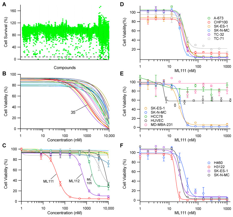
Ewing's sarcoma, characterized by pathognomonic t (11; 22) (q24; q12) and related chromosomal ETS family translocations, is a rare aggressive cancer of bone and soft tissue. Current protocols that include cytotoxic chemotherapeutic agents effectively treat localized disease; however, these aggressive therapies may result in treatment-related morbidities including second-site cancers in survivors. Moreover, the five-year survival rate in patients with relapsed, recurrent, or metastatic disease is less than 30%, despite intensive therapy with these cytotoxic agents. By using high-throughput phenotypic screening of small molecule libraries, we identified a previously uncharacterized compound (ML111) that inhibited in vitro proliferation of six established Ewing's sarcoma cell lines with nanomolar potency. Proteomic studies show that ML111 treatment induced prometaphase arrest followed by rapid caspase-dependent apoptotic cell death in Ewing's sarcoma cell lines. ML111, delivered via methoxypoly(ethylene glycol)-polycaprolactone copolymer nanoparticles, induced dose-dependent inhibition of Ewing's sarcoma tumor growth in a murine xenograft model and invoked prometaphase arrest in vivo, consistent with in vitro data. These results suggest that ML111 represents a promising new drug lead for further preclinical studies and is a potential clinical development for the treatment of Ewing's sarcoma.

Keywords: Ewing’s sarcoma; ML111; cancer; chemotherapy; drug discovery; high-throughput screening; nanoparticle drug delivery.


Conflict of interest statement

The authors declare no conflict of interest.

Figures

Figure 1
Figure 1
Lead compound selection process and the results of each step. (A) Primary screen: A scatter plot of the percentage cell survival of SK-ES-1 treated with 10,500 pure compounds at 10 μM for 48 h. Each dot represents one compound. Fifty-two hits (0.5% of the screened chemicals) were selected for further studies based on their ability to reduce cell viability by 85% (the dashed line shows the cutoff for selection of hits). (B) Secondary screen: Confirming the activity of the identified hits from the primary screen. The dose–response curve studies confirmed the activity of twenty-seven (51.9%) compounds. Compound 35 was selected for the tertiary screen. (C) Tertiary screen: The dose–response analysis of compound 35 and its structural analogs. ML111 was selected as the lead compound for further studies. (D) Dose–response analysis of Ewing’s sarcoma cell lines with either type I or type II fusion protein after ML111 treatment. (E,F) Effect of ML111 on various cancerous and primary cell lines. Data in (CF) shown as mean ± SD (n = 3).
Scheme 1
Scheme 1
Potency of ML111 analogs. Scheme shows the structures and SK-ES-1 cell growth inhibitory activity as dose–response IC50 values.
Figure 2
Figure 2
Enantiospecificity of ML111 action in Ewing’s sarcoma cells. (A,B) (R)-ML111 and (S)-ML111 chemical structures. (C) The racemate ML111 was resolved into its individual enantiomers by chiral chromatography. HPLC analysis of racemate clearly identified two single enantiomer peaks. AU: absorbance units. (D) CD spectra of (R)-ML111, (S)-ML111, and racemate ML111. The first enantiomer to elute from the column in (C) shows a specific rotation value of −180°, while that for the second enantiomer is +180°. (E) Dose–response analysis of racemate ML111, (S)-ML111, and (R)-ML111 in SK-N-MC cells with various concentrations of each compound ranging from 0.001 to 1 μM for 48 h.
Figure 3
Figure 3
Induction of caspase-dependent apoptosis by ML111 in a time-dependent manner. (A) Induction of caspase 3/7 activity after vehicle, ML111 (50 nM), or cabozantinib (250 nM) treatment at indicated time intervals. Green nuclear fluorescence indicates cellular caspase 3/7 activation as detected by using CellEvent Caspase-3/7 Green Detection Reagent. (B) Quantification of caspase-3/7 activated cells at indicated times. The mean is shown, and the shaded regions represent the SEM (n = 4). (C) Immunoblot analysis of pro-caspase 3 cleavage forming active caspase 3 after 12 h of treatment with ML111. (D) Western blot analysis of PARP cleavage as a marker of caspase 3 activation and apoptosis. (E) Measurement of early and late apoptosis in ML111 treated cells as measured by annexin V and propidium iodide staining and flow cytometry. (F) Caspase inhibitor Z-VAD attenuates ML111-mediated induction of apoptosis. Asterisks (**) indicate statistically significant rescue of ML111 induced apoptosis by Z-VAD treatment (p < 0.01). GAPDH and α-tubulin were used as loading controls. CASP-3: Caspase 3; C-CASP3: Cleaved Caspase 3; C-PARP: Cleaved PARP. Scale bar in panel A = 200 µm.
Figure 4
Figure 4
ML111 induced prometaphase arrest without altering microtubule dynamics. (A) Cell cycle analyses by flow cytometry in asynchronous SK-N-MC cells treated with 0.1% DMSO (upper panel) or 100 nM ML111 (lower panel). (B) Immunoblot analysis for cyclin B1, p-H3Ser10, and CDC20 in lysates from asynchronized SK-N-MC cells treated with 100 nM ML111 or 0.1% DMSO. Total histone H3 (H3) antibody was used as a control. (C) Cell-free tubulin polymerization assay. ML111 neither stimulated nor inhibited tubulin polymerization in vitro. Paclitaxel and colchicine were used as tubulin polymerization inducer and inhibitor agents, respectively. General tubulin buffer (Cytoskeleton, Inc.) was used as a control. AU: absorbance units. (D) ML111 induced prometaphase arrest. Asynchronized SK-N-MC cells were treated with either DMSO (0.1%) or ML111 (25 nM) for 12 h. The nuclei were stained with DAPI (blue), microtubules (α-tubulin, red), and mitotic chromosomes (p-H3Ser10, green). One representative result was shown from three independent experiments. Magnification: 63×. Scale bar = 10 μm.
Figure 5
Figure 5
ML111-induced accumulation of APC/C substrates in SK-N-MC cells. (A) Scatter plot showing relative quantitative enrichment of 7122 proteins by mass spectrometry in response to ML111 relative to vehicle control in SK-N-MC cells. Known cell cycle proteins upregulated > 1.5-fold (log2 > 0.58) are shown in blue. (B) Diagram showing where the accumulating APC/C substrates would normally be ubiquitinated by the APC/C complex in the cell cycle. The diagram illustrates the dependence the cell cycle progression on APC/C co-activators CDC20 and CDH1, the spindle assembly checkpoint (SAC), mitotic checkpoint complex (MCC), and the location in the cell cycle where various substrates are expected to be ubiquitinated.
Figure 6
Figure 6
Characterization of ML111-NP. (A) Cryo-TEM image of ML111-NP. See Figure S2 for a full-resolution image. (B) ML111 released from nanoparticles in two phases in vitro. (C) Effect of empty NP and ML111-NP on viability of SK-N-MC and HEK293 cells after 48 h of incubation. (D) Fluorescence images of nude mice with subcutaneous SK-N-MC tumors pre-, post-, and at 1, 3, 6, 10, and 24 h after i.v. injection of ML111-NP (co-encapsulated with the NIR SiNc dye for fluorescence visualization) and fluorescence images of various organs and tumors at 24 h post-injection. Images were recorded on the Pearl Impulse Scheme 800 nm channel. (E) Representative mice bearing subcutaneous SK-N-MC xenografts and treated with empty nanoparticles or those containing ML111, as indicated. Mice are shown at necropsy. Reduction in xenograft tumor volume was observed in mice treated with ML111-NP. (F) A tumor growth profile after i.v. injections with ML111-NP, empty nanoparticles, or 5% dextrose. Data are shown as mean ± SD (n = 3). (G) Immunohistochemical staining with anti-p-H3Ser10 in SK-N-MC xenograft tissues from mice treated with empty nanoparticles or those loaded with ML111 (15 mg of ML111/kg). See Figure S6 for a quantification of the results. Scale bar = 20 nm in panel A and 100 µm in panel G.

References

    1. Smith M.A., Altekruse S.F., Adamson P.C., Reaman G.H., Seibel N.L. Declining Childhood and Adolescent Cancer Mortality. Cancer. 2014;120:2497–2506. doi: 10.1002/cncr.28748. - DOI - PMC - PubMed
    1. Riggi N., Suvà M.L., Stamenkovic I. Ewing’s Sarcoma. N. Engl. J. Med. 2021;384:154–164. doi: 10.1056/NEJMra2028910. - DOI - PubMed
    1. Bailey K., Cost C., Davis I., Glade-Bender J., Grohar P., Houghton P., Isakoff M., Stewart E., Laack N., Yustein J., et al. Emerging Novel Agents for Patients with Advanced Ewing Sarcoma: A Report from the Children’s Oncology Group (COG) New Agents for Ewing Sarcoma Task Force. F1000Research. 2019;8:493. doi: 10.12688/f1000research.18139.1. - DOI - PMC - PubMed
    1. Womer R.B., West D.C., Krailo M.D., Dickman P.S., Pawel B.R., Grier H.E., Marcus K., Sailer S., Healey J.H., Dormans J.P., et al. Randomized Controlled Trial of Interval-Compressed Chemotherapy for the Treatment of Localized Ewing Sarcoma: A Report from the Children’s Oncology Group. J. Clin. Oncol. 2012;30:4148–4154. doi: 10.1200/JCO.2011.41.5703. - DOI - PMC - PubMed
    1. Lessnick S.L., Braun B.S., Denny C.T., May W.A. Multiple Domains Mediate Transformation by the Ewing’s Sarcoma EWS/FLI-1 Fusion Gene. Oncogene. 1995;10:423–431. - PubMed

LinkOut - more resources