. 2021 Sep 30;13(10):1592.
doi: 10.3390/pharmaceutics13101592.

Bacterial Cellulose as a Potential Bio-Scaffold for Effective Re-Epithelialization Therapy

Affiliations

Bacterial Cellulose as a Potential Bio-Scaffold for Effective Re-Epithelialization Therapy

Juin-Hong Cherng et al. Pharmaceutics. .

Abstract

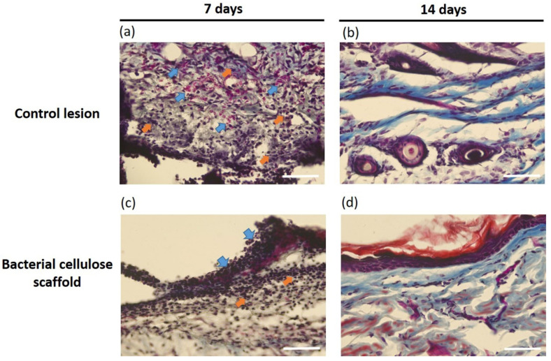
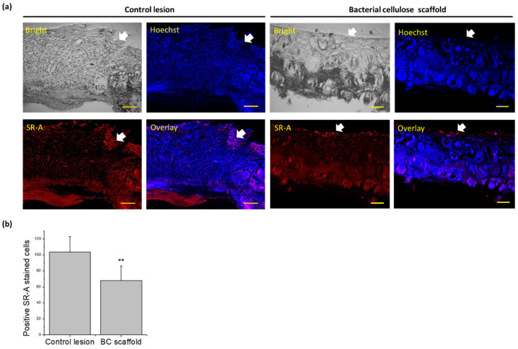
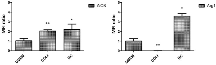
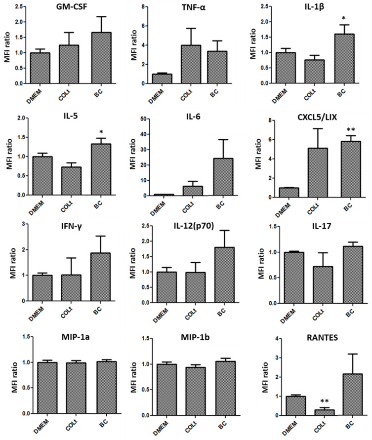
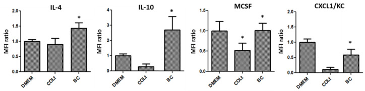
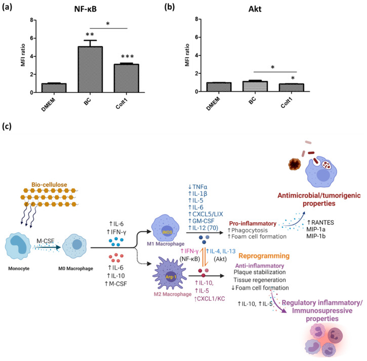
Currently, there are several therapeutic approaches available for wound injury management. However, a better understanding of the underlying mechanisms of how biomaterials affect cell behavior is needed to develop potential repair strategies. Bacterial cellulose (BC) is a bacteria-produced biopolymer with several advantageous qualities for skin tissue engineering. The aim here was to investigate BC-based scaffold on epithelial regeneration and wound healing by examining its effects on the expression of scavenger receptor-A (SR-A) and underlying macrophage behavior. Full-thickness skin wounds were generated on Sprague-Dawley rats and the healing of these wounds, with and without BC scaffolds, was examined over 14 days using Masson's trichome staining. BC scaffolds displayed excellent in vitro biocompatibility, maintained the stemness function of cells and promoted keratinocyte differentiation of cells, which are vital in maintaining and restoring the injured epidermis. BC scaffolds also exhibited positive in vivo effects on the wound microenvironment, including improved skin extracellular matrix deposition and controlled excessive inflammation by reduction of SR-A expression. Furthermore, BC scaffold significantly enhanced epithelialization by stimulating the balance of M1/M2 macrophage re-programming for beneficial tissue repair relative to that of collagen material. These findings suggest that BC-based materials are promising products for skin injury repair.

Keywords: bacterial cellulose; epithelialization; scaffold; tissue regeneration; wound healing.


Conflict of interest statement

The authors declare no conflict of interest.

Figures

Figure 1
Figure 1
Characteristics of bacterial cellulose scaffold. (a) Scanning electron microscopy. (b) Fourier-transform infrared spectroscopic analysis.
Figure 2
Figure 2
Study of in vitro biocompatibility of bacterial cellulose scaffold incubated with human adipose stem cells (hASCs) using trypan blue staining. (a) Growth of hASCs with time. (b) Quantification of cell survival (scale bar = 200 µm; * p < 0.05).
Figure 3
Figure 3
Immunofluorescence staining of human adipose stem cells cultured on bacterial cellulose scaffold for 14 days. (a,b) Octamer-binding protein 4 (OCT-4) and OCT-4/Hoechst overlay expression, respectively. (c,d) Nestin and Nestin/Hoechst overlay expression, respectively. (e,f) Sex determining region Y-box 9 (SOX-9) and SOX-9/Hoechst overlay expression, respectively (scale bar = 200 µm).
Figure 4
Figure 4
Rat model of skin defects healing over time without (left side) or with bacterial cellulose scaffold covering (right side).
Figure 5
Figure 5
Histological analysis of rat skin defects using Masson’s trichrome staining. (a,b) Control lesion group after 7 and 14 days of treatment, respectively. (c,d) Bacterial cellulose scaffold group after 7 and 14 days of treatment, respectively (blue arrow = immune cell infiltration; orange arrow = granulation tissue; scale bar = 50 µm).
Figure 6
Figure 6
Immunostaining of scavenger receptor-A (SR-A) expression in rat skin defects after 7 days of treatment. (a) Control lesion compared with bacterial cellulose (BC) scaffold. (b) Semi-quantitative analysis of SR-A positively-stained cells using ImageJ (scale bar = 200 µm; ** p < 0.01).
Figure 7
Figure 7
Expression of inducible nitric oxide synthase (iNOS) and arginase-1 (Arg1) secreted by macrophages cultured on bacterial cellulose (BC) scaffold, collagen total type material (COLt), or in DMEM for 24 h (* p < 0.05; ** p < 0.01).
Figure 8
Figure 8
Expression of M1 macrophage-related cytokines secreted by macrophages cultured on bacterial cellulose (BC) scaffold, collagen total type material (COLt), or in DMEM for 24 h (GM-CSF = granulocyte-macrophage colony-stimulating factor; TNF-α = tumor necrosis factor-alpha; IL-1β = interleukin-1 beta; IL-5 = interleukin-5; IL-6 = interleukin-6; CXCL5/LIX = chemokine (C-X-C motif) ligand 5/lipopolysaccharide-induced chemokine; IFN-γ = interferon-gamma; IL-12(p70) = interleukin-12(p70); IL-17 = interleukin-17; MIP1a = macrophage inflammatory protein-1 alpha; MIP1b = macrophage inflammatory protein-1 beta; RANTES = regulated on activation, normal t expressed and secreted; * p < 0.05; ** p < 0.01).
Figure 9
Figure 9
Expression of M2 macrophage-related cytokines secreted by macrophages cultured on bacterial cellulose (BC) scaffold, collagen total type material (COLt), or in DMEM for 24 h (IL-4 = interleukin-4; IL-10 = interleukin-10; MCSF = macrophage colony-stimulating factor; CXCL1/KC = chemokine (C-X-C motif) ligand 1/keratinocyte-derived chemokine; * p < 0.05).
Figure 10
Figure 10
Signaling pathways alteration in macrophages affected by bacterial cellulose (BC) scaffold after 24 h of treatment. (a) Nuclear factor kappa B (NF-κB). (b) Protein kinase B (Akt). (c) Summary of macrophage polarization states (this figure was created with BioRender.com and based on icons from biorender.com). (* p < 0.05; ** p < 0.01; *** p < 0.001).

Similar articles

Cited by

References

    1. Das S., Baker A.B. Biomaterials and nanotherapeutics for enhancing skin wound healing. Front. Bioeng. Biotechnol. 2016;4:82. doi: 10.3389/fbioe.2016.00082. - DOI - PMC - PubMed
    1. Vig K., Chaudhari A., Tripathi S., Dixit S., Sahu R., Pillai S., Dennis V.A., Singh S.R. Advances in skin regeneration using tissue engineering. Int. J. Mol. Sci. 2017;18:789. doi: 10.3390/ijms18040789. - DOI - PMC - PubMed
    1. Debels H., Hamdi M., Abberton K., Morrison W. Dermal matrices and bioengineered skin substitutes: A critical review of current opinions. Plast. Reconstr. Surg. Glob. Open. 2015;3:e284. doi: 10.1097/GOX.0000000000000219. - DOI - PMC - PubMed
    1. Mitchell A.C., Briquez P.S., Hubbell J.A., Cochran J.R. Engineering growth factors for regenerative medicine applications. Acta Biomater. 2016;30:1–12. doi: 10.1016/j.actbio.2015.11.007. - DOI - PMC - PubMed
    1. Yu J.R., Navarro J., Coburn J.C., Mahadik B., Molnar J., Holmes J.H., Nam A.J., Fisher J.P. Current and future perspectives on skin tissue engineering: Key features of biomedical research, translational assessment, and clinical application. Adv. Healthc. Mater. 2019;8:1801471. doi: 10.1002/adhm.201801471. - DOI - PMC - PubMed