. 2021 Oct 3;13(10):1608.
doi: 10.3390/pharmaceutics13101608.

Human Skin Permeation Enhancement Using PLGA Nanoparticles Is Mediated by Local pH Changes

Affiliations

Human Skin Permeation Enhancement Using PLGA Nanoparticles Is Mediated by Local pH Changes

Javiana Luengo et al. Pharmaceutics. .

Abstract

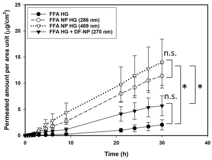
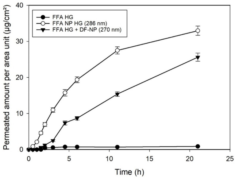
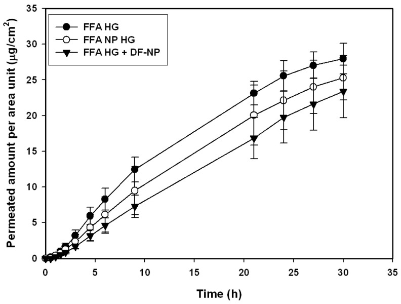
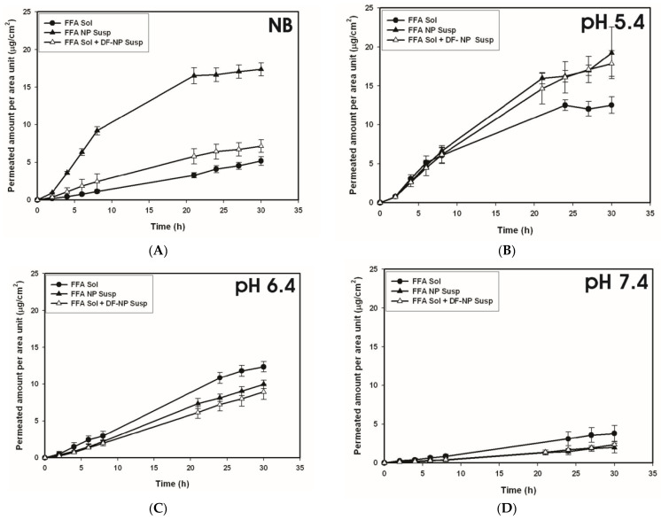
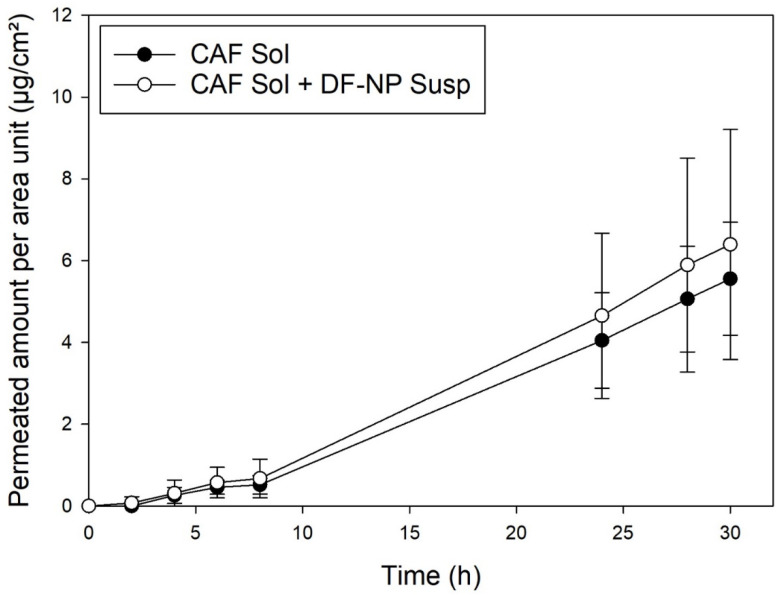
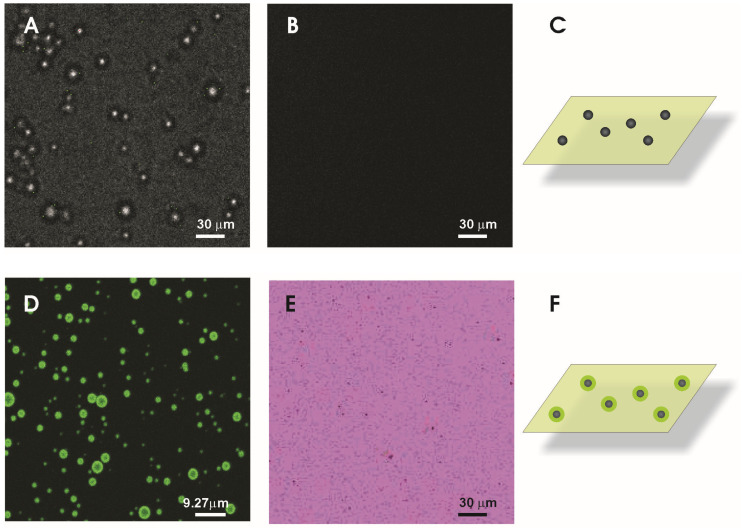
The steady improvement and optimization of transdermal permeation is a constant and challenging pharmaceutical task. In this study the influence of poly(lactide-co-glycolide) (PLGA) nanoparticles on the dermal permeation of the anti-inflammatory drug flufenamic acid (FFA) was investigated. For this aim, different vehicles under non-buffered and buffered conditions and different skin models (human heat separated epidermis and reconstructed human epidermis equivalents) were tested. Permeation experiments were performed using static Franz diffusion cells under infinite dosing conditions. Already the presence of drug-free nanoparticles increased drug permeation across the skin. Drug permeation was even enhanced when applying drug-loaded nanoparticles. In contrast, buffered vehicles with different pH values (pH 5.4-7.4) revealed the influence of the pH on the permeation of FFA. The change of the surrounding pH of the biodegradable nanoparticulate system was demonstrated and visualized using pH-sensitive fluorescent probes. While a potential contribution of hair follicles could be ruled out, our data suggest that the enhanced permeation of FFA through human skin in the presence of PLGA nanoparticles is mediated by a locally decreased pH during hydrolytic degradation of this polymer. This hypothesis is supported by the observation that skin permeation of the weak base caffeine was not affected.

Keywords: PLGA; human skin; pH effects; penetration; permeation; polymer nanoparticles.


Conflict of interest statement

The authors declare no conflict of interest.

Figures

Figure 1
Figure 1
Permeation profiles across heat separated human epidermis of flufenamic acid using infinite dose regime of non-buffered hydrogels containing: dissolved drug (FFA HG), FFA-loaded nanoparticles of different sizes (FFA NP HG), and dissolved drug with drug-free nanoparticles (FFA HG + DF-NP). Concentration 0.125 µg/g. The drug-loaded NP formulations were statistically not different (n.s.) from each other, but both were statistically different (*) from the drug-free formulation and the drug in hydrogel. Statistical evaluation is based on Papp values from 4.5 to 30 h.
Figure 2
Figure 2
Permeation profiles of reconstructed human epidermis (SkinEthic®) using flufenamic acid at infinite dose regime of: dissolved drug in hydrogel (FFA HG), FFA-loaded nanoparticles dispersed in the hydrogel (FFA NP HG), and dissolved drug with drug-free nanoparticles dispersed in the hydrogel (FFA HG + DF-NP). Concentration of FFA 0.125 µg/g.
Figure 3
Figure 3
Permeation profiles of human heat separated epidermis of a buffered hydrogel (pH 6.0) using infinite dose regime of flufenamic acid preparations as dissolved drug (FFA HG), FFA-loaded nanoparticles (FFA NP HG), and dissolved drug with drug-free nanoparticles (FFA HG + DF-NP).
Figure 4
Figure 4
Permeation profiles obtained for human heat separated epidermis using flufenamic acid solution (FFA sol), FFA solution plus drug-free nanoparticles (FFA sol + DF-NP susp) and drug-loaded nanoparticles (FFA NP susp). The formulations were applied as non-buffered preparations (NB, (A)) and buffered preparations at different pH values (pH 5.4 (B), pH6.4 (C), pH 7.4 (D)). Concentration of flufenamic acid 37.6 ± 1.9 µg/mL.
Figure 5
Figure 5
Permeation with heat separated epidermis: caffeine in solution (black, CAF Sol) and caffeine with drug-free PLGA nanoparticles (open symbols, CAF Sol + DF-NP). Caffeine concentration 100 µg/mL.
Figure 6
Figure 6
Fluorescence micrographs obtained after dispersing different particle fractions into pH sensor solution. Transmission light image were taken to identify Arabic gum/gelatine A microparticles (A) and the corresponding fluorescence image (B). PLGA microparticles show green fluorescence localized at the surface of the particles or the close surrounding (D). For nanoparticles, fluorescent spots were detected (E). For highlighting the particles position the color coding was inverted leading to a pink background with block spots representing the fluorescent nanoparticles. The sketches should illustrate the experimental conditions (C,F).

References

    1. Rodríguez Cruz I., Domínguez-Delgado C., Escobar-Chávez J., López-Cervantes M., Díaz-Torres R. Current Technologies to Increase the Transdermal Delivery of Drugs. Volume 2. Bentham Science Publishers; Sharjah, United Arab Emirates: 2016. Physical Penetration Enhancers: An Overview; pp. 3–34.
    1. Wang F.-Y., Chen Y., Huang Y.-Y., Cheng C.-M. Transdermal drug delivery systems for fighting common viral infectious diseases. Drug Deliv. Transl. Res. 2021;11:1498–1508. doi: 10.1007/s13346-021-01004-6. - DOI - PMC - PubMed
    1. Mota A.H., Rijo P., Molpeceres J., Reis C.P. Broad overview of engineering of functional nanosystems for skin delivery. Int. J. Pharm. 2017;532:710–728. doi: 10.1016/j.ijpharm.2017.07.078. - DOI - PubMed
    1. Palmer B.C., DeLouise L.A. Nanoparticle-Enabled Transdermal Drug Delivery Systems for Enhanced Dose Control and Tissue Targeting. Molecules. 2016;21:1719. doi: 10.3390/molecules21121719. - DOI - PMC - PubMed
    1. Prow T.W., Grice J.E., Lin L.L., Faye R., Butler M., Becker W., Wurm E.M.T., Yoong C., Robertson T.A., Soyer H.P., et al. Nanoparticles and microparticles for skin drug delivery. Adv. Drug Deliv. Rev. 2011;63:470–491. doi: 10.1016/j.addr.2011.01.012. - DOI - PubMed

LinkOut - more resources