. 2021 Oct 12;13(10):1663.
doi: 10.3390/pharmaceutics13101663.

Enhanced Detection of Desmoplasia by Targeted Delivery of Iron Oxide Nanoparticles to the Tumour-Specific Extracellular Matrix

Affiliations

Enhanced Detection of Desmoplasia by Targeted Delivery of Iron Oxide Nanoparticles to the Tumour-Specific Extracellular Matrix

Meenu Chopra et al. Pharmaceutics. .

Abstract

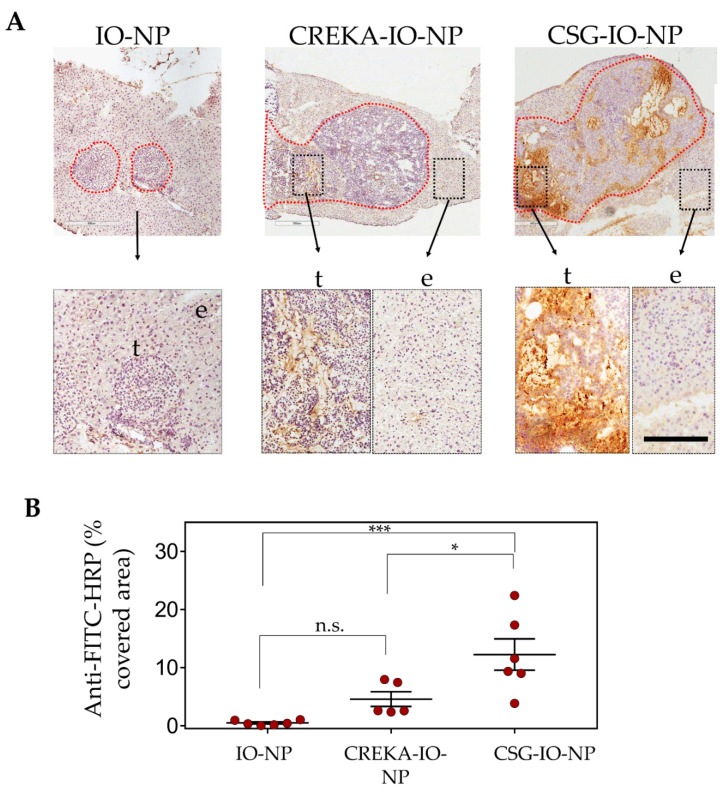
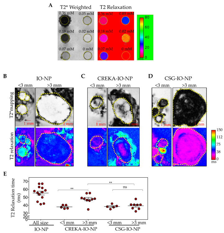
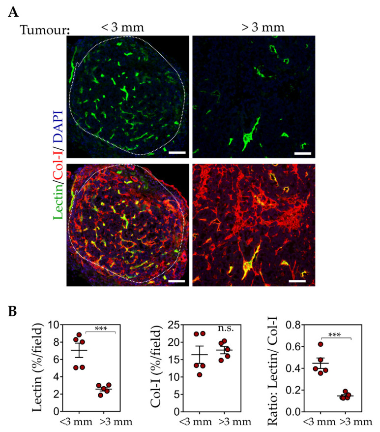
Diagnostic imaging of aggressive cancer with a high stroma content may benefit from the use of imaging contrast agents targeted with peptides that have high binding affinity to the extracellular matrix (ECM). In this study, we report the use of superparamagnetic iron-oxide nanoparticles (IO-NP) conjugated to a nonapeptide, CSGRRSSKC (CSG), which specifically binds to the laminin-nidogen-1 complex in tumours. We show that CSG-IO-NP accumulate in tumours, predominantly in the tumour ECM, following intravenous injection into a murine model of pancreatic neuroendocrine tumour (PNET). In contrast, a control untargeted IO-NP consistently show poor tumour uptake, and IO-NP conjugated to a pentapeptide. CREKA that bind fibrin clots in blood vessels show restricted uptake in the angiogenic vessels of the tumours. CSG-IO-NP show three-fold higher intratumoral accumulation compared to CREKA-IO-NP. Magnetic resonance imaging (MRI) T2-weighted scans and T2 relaxation times indicate significant uptake of CSG-IO-NP irrespective of tumour size, whereas the uptake of CREKA-IO-NP is only consistent in small tumours of less than 3 mm in diameter. Larger tumours with significantly reduced tumour blood vessels show a lack of CREKA-IO-NP uptake. Our data suggest CSG-IO-NP are particularly useful for detecting stroma in early and advanced solid tumours.

Keywords: CSG; extracellular matrix; magnetic resonance imaging; nanoparticles; tumour targeting.


Conflict of interest statement

The authors declare no conflict of interest. The funders had no role in the design of the study; in the collection, analyses, or interpretation of data; in the writing of the manuscript, or in the decision to publish the results.

Figures

Figure 1
Figure 1
CSG and CREKA specifically recognise different compartments in RIP1-Tag5 tumours. Tumour-bearing RIP1-Tag5 mice were i.v. injected with 0.1 µmol of FAM-CREKA or FAM-CSG, and tissues were collected after 1 h of circulation. (AD) show distribution of FAM-CREKA and FAM-CSG (green) in tumours co-stained either with the vascular marker CD31 or the ECM marker laminin (red). Scale bars: 50 μm.
Figure 2
Figure 2
Structural and morphological characterisation of IO-NPs. (A) High-resolution transmission electron microscopy (HR-TEM) micrographs of IO-NPs and (B) a magnified view of a single NP. (C) Selected area electron diffraction (SAED) pattern acquired from an IO-NP assembly.
Figure 3
Figure 3
Schematic illustration of IO-NP consisting of a dextran-coated IO particle (core, gray) encapsulated by DSPE-PEG2000 lipids (in red) with the lipids tagged to a peptide or cysteine residue (in green). FAM-X- Cys-CSG (CSGRRSSKC), FAM-X-CREKA or FAM-X-Cys (untargeted) is tagged to the lipids via cysteine-maleimide (Mal) interaction (shown in the box).
Figure 4
Figure 4
CSG and CREKA deliver IO-NP to a specific target in solid tumours. Mice bearing RIP1-Tag5 tumours were i.v. injected with 100 μL of the indicated IO-NP (5 mg/kg Fe), and tissues were collected after 4 h of circulation. (A) Top: Representative micrographs of tumours (circled, red) with surrounding exocrine pancreas. Nanoparticle uptake in these tissues was detected based on anti-FITC-HRP reactivity (brown) and nuclei were counterstained with haematoxylin. Scale bar, 200 µm. Bottom: Higher magnification of the tumour area (t) and exocrine pancreas (e). (B) Plots of % area per tumour that was positive for anti-FITC antibody detection (in A) for tumours with untargeted-IO-NP, CREKA-IO-NP and CSG-IO-NP, and mean ± SEM (n = 3 mice/group *** p < 0.005, * p < 0.05, n.s. = not significant, by one-way ANOVA test).
Figure 5
Figure 5
RIP1-Tag5 tumours with (A) CREKA-IO-NP and (B) CSG-IO-NP (green) were co-stained with CD31 (red), collagen I (Col-1, red), laminin (red) or nidogen-1 (red), and nuclei were stained with DAPI (blue). Representative micrographs are shown with indicated detection of IO-NP accumulation (arrows). Scale bars, 50 µm.
Figure 6
Figure 6
CSG-IO-NP show greater MRI tumour contrast compared to untargeted or CREKA-IO-NP. (A) T2*-weighted images (black and white) and T2 relaxation maps (coloured) of IO-NP at indicated concentrations ranging from 0 to 0.36 mM Fe. (BD) Representative images of T2*-weighted (top) and T2 relaxation maps (magenta, bottom) of tumours with exocrine pancreas following ex vivo MRI scan. Tumours were analysed based on their sizes (<3 mm or >3mm in diameter). Scale bar: 1 mm. RIP1-Tag5 tumours and exocrine tissue were isolated after 4 h of in vivo circulation of untargeted-IO-NP, CREKA-IO-NP and CSG-IO-NP in tumour-bearing mice (n = 10–15 tumours from n = 5–6 mice per group, 5 mg/kg Fe). Mice were perfused with PBS to remove the unbound particles. The MRI scan was performed on formalin-fixed tumours embedded in 2% agarose. (E) Reduction in T2 relaxation time indicates the relative increase of IONP in individual tumours and mean ± SEM (** p < 0.01 by Student’s t-test. n.s. = not significant).
Figure 7
Figure 7
ECM and blood vessel contents in tumours correlate with the effectiveness of CSG and CREKA to deliver IO-NP. (A) Lectin+ vessels (green) in tumours after RIP1-Tag5 mice received an i.v. injection of FITC-labelled lectin (0.1 μmol). Top: Representative micrographs show % of lectin+ vessels in small tumours (diameter < 3 mm) and larger tumours (diameter > 3 mm). Bottom: The same micrographs indicating lectin+ area and collagen I expression (red). Scale bars: 100 μm. (B) Quantification of % lectin+/tumour (left), % collagen-I+ expression/tumour (middle) and ratio of lectin:collagen-I (right) with mean ± SEM are shown (10 tumours from n = 3 RIP1-Tag5 mice (*** p < 0.001 for Student’s t-test).

References

    1. Schneble E.J., Graham L.J., Shupe M.P., Flynt F.L., Banks K.P., Kirkpatrick A.D., Nissan A., Henry L., Stojadinovic A., Shumway N.M., et al. Current approaches and challenges in early detection of breast cancer recurrence. J. Cancer. 2014;5:281–290. doi: 10.7150/jca.8016. - DOI - PMC - PubMed
    1. Singhi A.D., Koay E.J., Chari S.T., Maitra A. Early detection of pancreatic cancer: Opportunities and challenges. Gastroenterology. 2019;156:2024–2040. doi: 10.1053/j.gastro.2019.01.259. - DOI - PMC - PubMed
    1. Zhang L., Sanagapalli S., Stoita A. Challenges in diagnosis of pancreatic cancer. World J. Gastroenterol. 2018;24:2047–2060. doi: 10.3748/wjg.v24.i19.2047. - DOI - PMC - PubMed
    1. Chi C., Du Y., Ye J., Kou D., Qiu J., Wang J., Tian J., Chen X. Intraoperative imaging-guided cancer surgery: From current fluorescence molecular imaging methods to future multi-modality imaging technology. Theranostics. 2014;4:1072–1084. doi: 10.7150/thno.9899. - DOI - PMC - PubMed
    1. Madajewski B., Judy B.F., Mouchli A., Kapoor V., Holt D., Wang M.D., Nie S., Singhal S. Intraoperative near-infrared imaging of surgical wounds after tumor resections can detect residual disease. Clin. Cancer Res. 2012;18:5741–5751. doi: 10.1158/1078-0432.CCR-12-1188. - DOI - PMC - PubMed

LinkOut - more resources