. 2021 Oct 18;13(10):1724.
doi: 10.3390/pharmaceutics13101724.

Design of a Platelet-Mediated Delivery System for Drug-Incorporated Nanospheres to Enhance Anti-Tumor Therapeutic Effect

Affiliations

Design of a Platelet-Mediated Delivery System for Drug-Incorporated Nanospheres to Enhance Anti-Tumor Therapeutic Effect

Jun-Ichiro Jo et al. Pharmaceutics. .

Abstract

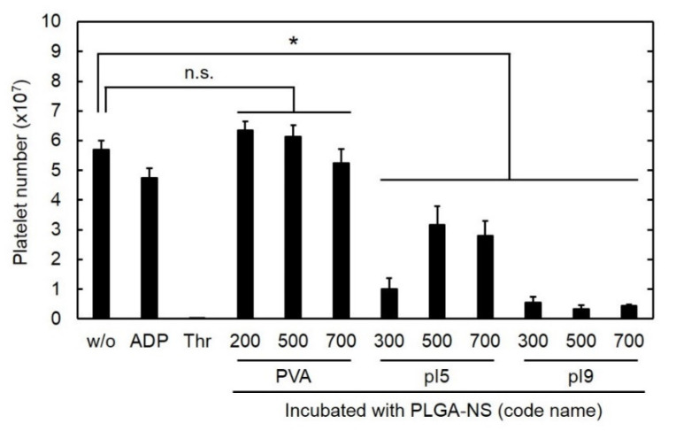
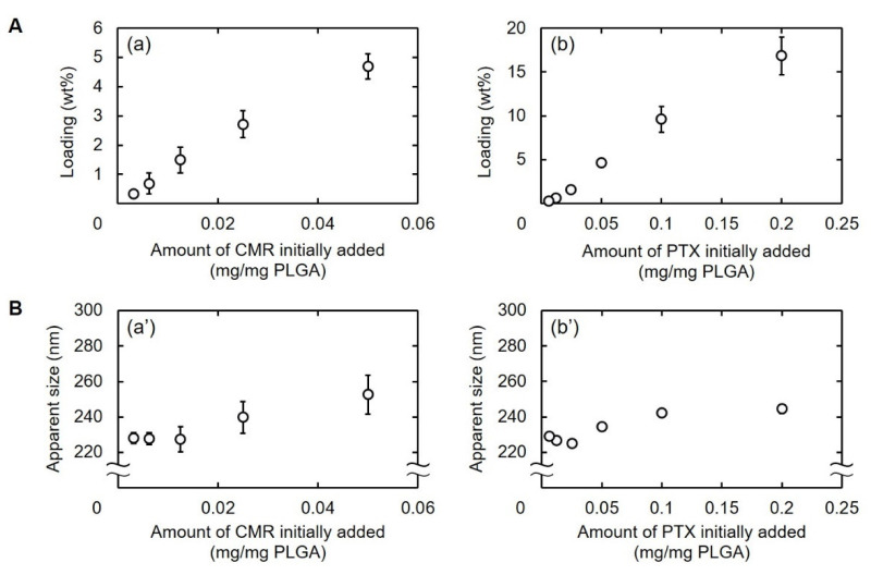
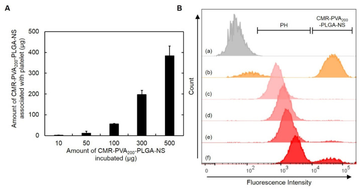
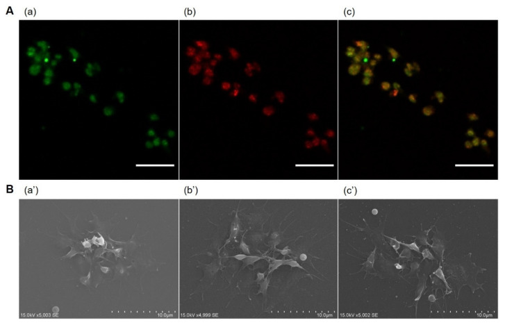
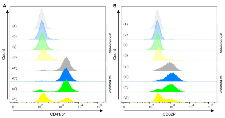
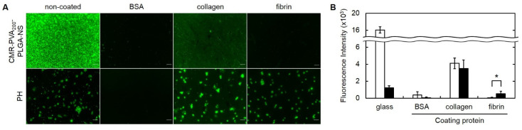
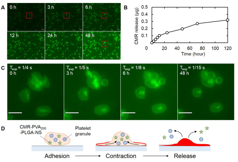
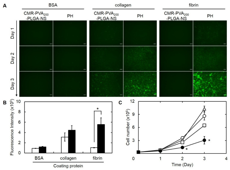
The objective of this study is to construct a platelet-mediated delivery system for drug-incorporated nanospheres. Nanospheres of poly(lactic-co-glycolic acid) (PLGA-NS) with different sizes and surface properties were prepared by changing the preparation parameters, such as the type of polymer surfactant, the concentration of polymer surfactant and PLGA, and the stirring rate. When incubated with platelets, PLGA-NS prepared with poly(vinyl alcohol) suppressed the platelet activation. Scanning electron microscopic and flow cytometry examinations revealed that platelets associated with PLGA-NS (platelet hybrids, PH) had a similar appearance and biological properties to those of the original platelets. In addition, the PH with PLGA-NS specifically adhered onto the substrate pre-coated with fibrin to a significantly great extent compared with PLGA-NS alone. When applied in an in vitro model of tumor tissue which was composed of an upper chamber pre-coated with fibrin and a lower chamber culturing tumor cells, the PH with PLGA-NS incorporating an anti-tumor drug were delivered to the tumor cells through the specific adhesion onto the upper chamber and, consequently, drug release from the upper chamber took place, resulting in the growth suppression of tumor cells. It is concluded that the drug delivery system based on PH is promising for tumor treatment.

Keywords: drug delivery system; nanopsheres; platelets; poly(lactic-co-glycolic acid); tumor.


Conflict of interest statement

The authors declare no conflict of interest.

Figures

Figure 1
Figure 1
Number of platelets 30 h after incubation with PLGA-NS of different sizes prepared in the presence of various surfactants (PVA, pI5, and pI9). The ADP and thrombin (Thr) were used as agonists for platelet activation. n.s.: not significant. * p < 0.05: significant differences between two groups.
Figure 2
Figure 2
(A) Loading percentages of CMR (a) or PTX into PLGA-NS (b). (B) Apparent sizes of PVA200-PLGA-NS incorporating CMR (a’) or PTX (b’). Various amounts of CMR or PTX were initially added for nanospheres preparation.
Figure 3
Figure 3
(A) The amount of CMR-PVA200-PLGA-NS associated with platelets 30 min after incubation with different amounts of CMR-PVA200-PLGA-NS. (B) Flow cytometric histograms of platelets (a), CMR-PVA200-PLGA-NS (b), or PH obtained by incubation with 10 (c), 50 (d), 100 (e), or 300 µg of CMR-PVA200-PLGA-NS (f).
Figure 4
Figure 4
(A) Confocal microscopic images of PH incorporating CMR-PVA200-PLGA-NS. The PH were visualized by (a) CMR (green) incorporated into PVA200-PLGA-NS and (b) the staining with anti-CD41/61 antibody (red), then the merged image was obtained (c). Scale bar is 10 µm. (B) Scanning electron microscopic images of platelets (a’) or PH prepared by the incubation with PVA200-PLGA-NS (b’) or PTX-PVA200-PLGA-NS (c’) aiming at the observation of aggregating behavior on the glass bottom dish.
Figure 5
Figure 5
Flow cytometric histograms of platelets (a), PH with PVA200-PLGA-NS (b) or PTX-PVA200-PLGA-NS (c), or platelets incubated with PTX (d) under the treatment without (blurred colors, a–d) or with thrombin (plain colors, a’–d’). The platelets or PH were stained with CD41/61 (A) or CD62P (B). The amount of PTX in the PH (c, c’) was same as that incubated with platelets (d, d’).
Figure 6
Figure 6
(A) Fluorescent microscopic images of CMR-PVA200-PLGA-NS or their PH adhered on the bare glass bottom dish or the dish pre-coated with BSA, collagen, and fibrin. (B) The fluorescence intensity 30 min after adding CMR-PVA200-PLGA-NS (□) or their PH adhered on the bare glass bottom dish or the dish pre-coated with proteins (BSA, collagen, or fibrin) (■). Six images were analyzed by using ImageJ 1.53. * p < 0.05: significant difference between two groups.
Figure 7
Figure 7
Time course of CMR-PVA200-PLGA-NS released from PH. (A) Time-lapse fluorescent microscopic images 0, 3, 6, 12, 24, and 48 h after incubation of PH. Scale bar is 10 µm. (B) Time profile of CMR release from PH. (C) Enlarged fluorescent microscopic images of red frame boxes indicated in (A). The values of Texp indicates exposure time in the corresponding incubation periods. Scale bar is 5 µm. (D) The scheme of plausible mechanism on the release of CMR-PVA200-PLGA-NS from PH.
Figure 8
Figure 8
(A) Fluorescence microscopic images of B16F10 melanoma cells during 3 days of culture on the lower chamber inserted with the upper Transwell® chamber pre-coated with BSA, collagen, or fibrin, followed by the culture with CMR-PVA200-PLGA-NS or their PH on day 0. Scale bar is 100 µm. (B) Fluorescence intensity of CMR in B16F10 cells 3 days after culture on the lower chamber inserted with the upper Transwell® chamber pre-coated with BSA, collagen, or fibrin, followed by the culture with CMR-PVA200-PLGA-NS (□) or their PH on day 0 (■). * p < 0.05: significant difference between two groups. (C) Time profiles of B16F10 cells number cultured on the lower chamber inserted with the upper Transwell® chamber pre-coated with fibrin, followed by the culture without (○) or with platelet (△), PTX-PVA200-PLGA-NS (□), and their PH on day 0 (●). The amount of PTX incorporated in each group (PTX-PVA200-PLGA-NS or PH) is 5.5 μg. * p < 0.05: significant difference against other groups at the corresponding time.

Similar articles

Cited by

References

    1. Aparicio-Blanco J., Sanz-Arriazu L., Lorenzoni R., Blanco-Prieto M.J. Glioblastoma chemotherapeutic agents used in the clinical setting and in clinical trials: Nanomedicine approaches to improve their efficacy. Int. J. Pharm. 2020;581:119283. doi: 10.1016/j.ijpharm.2020.119283. - DOI - PubMed
    1. Dhupal M., Chowdhury D. Phytochemical-Based Nanomedicine for Advanced Cancer Theranostics: Perspectives on Clinical Trials to Clinical Use. Int. J. Nanomed. 2020;15:9125–9157. doi: 10.2147/IJN.S259628. - DOI - PMC - PubMed
    1. Fraguas-Sanchez A.I., Martin-Sabroso C., Fernandez-Carballido A., Torres-Suarez A.I. Current status of nanomedicine in the chemotherapy of breast cancer. Cancer Chemother. Pharmacol. 2019;84:689–706. doi: 10.1007/s00280-019-03910-6. - DOI - PubMed
    1. Majumder J., Taratula O., Minko T. Nanocarrier-based systems for targeted and site specific therapeutic delivery. Adv. Drug. Deliv. Rev. 2019;144:57–77. doi: 10.1016/j.addr.2019.07.010. - DOI - PMC - PubMed
    1. Cai S.S., Li T., Akinade T., Zhu Y., Leong K.W. Drug delivery carriers with therapeutic functions. Adv. Drug Deliv. Rev. 2021;176:113884. doi: 10.1016/j.addr.2021.113884. - DOI - PMC - PubMed

LinkOut - more resources