. 2021 Sep 26;10(10):1245.
doi: 10.3390/pathogens10101245.

Genetic Variability and Evidence of a New Subgroup in Watermelon Mosaic Virus Isolates

Affiliations

Genetic Variability and Evidence of a New Subgroup in Watermelon Mosaic Virus Isolates

Osama A Abdalla et al. Pathogens. .

Abstract

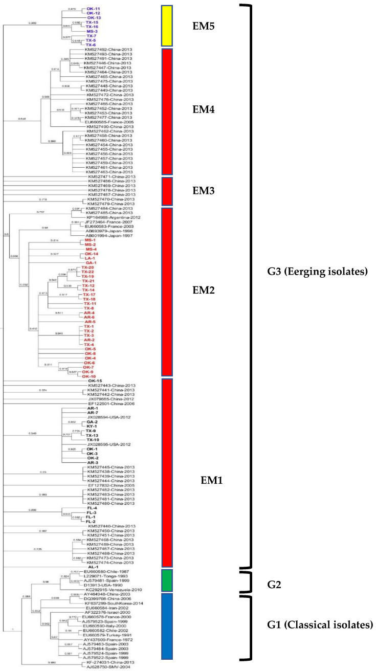
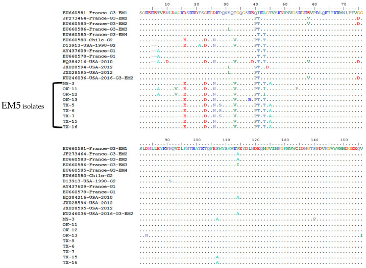
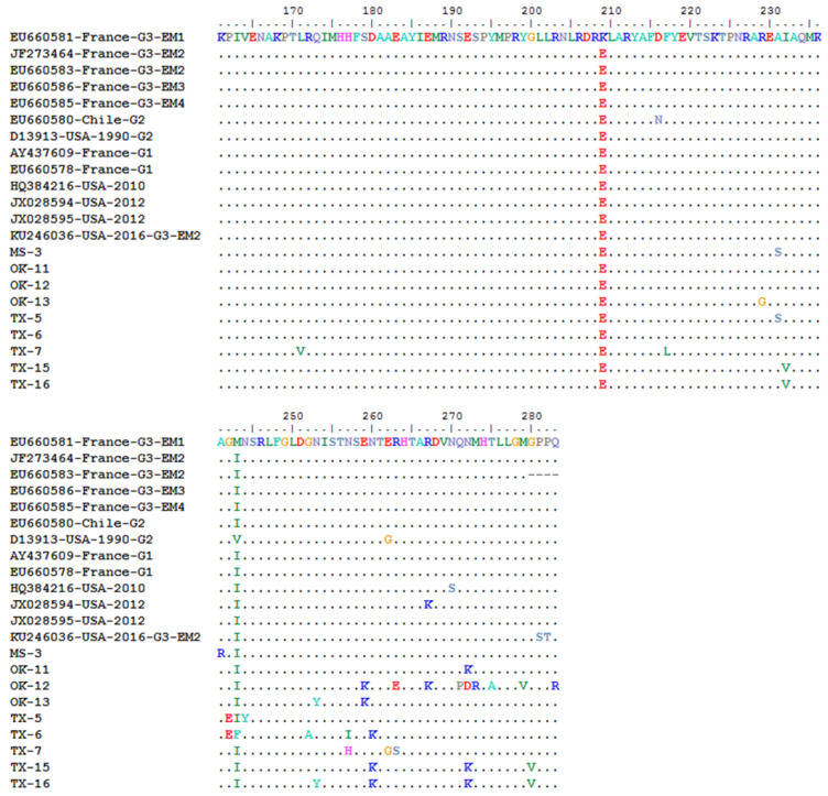
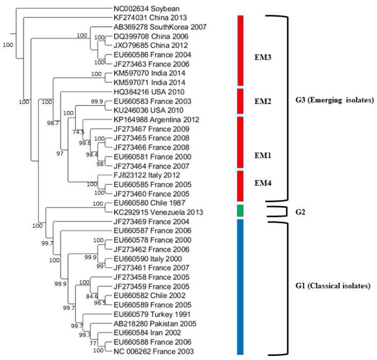
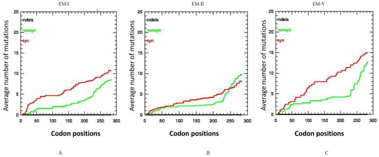
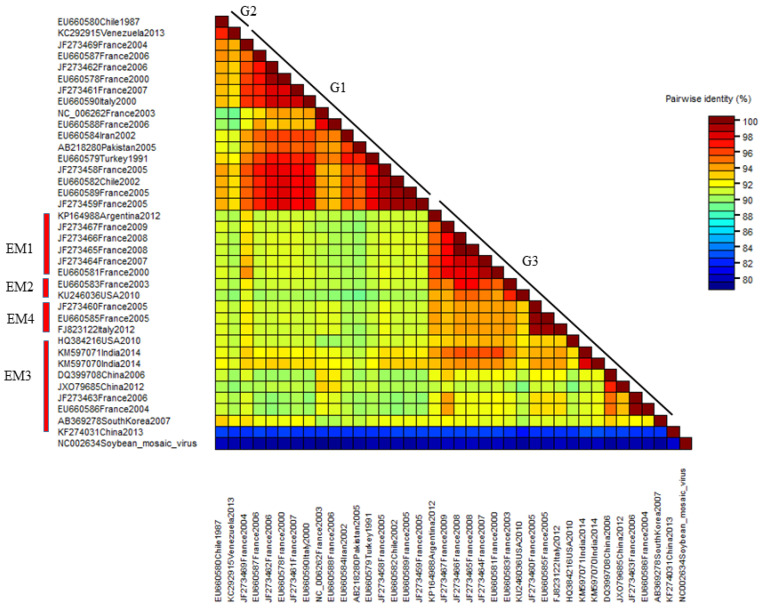
Watermelon mosaic virus (WMV) is one of the important Potyviruses that infect cucurbits worldwide. To better understand the population structure of WMV in the United States (U.S.), 57 isolates were collected from cucurbit fields located in nine southern states. The complete coat protein gene of all WMV isolates was cloned, sequenced and compared with 89 reported WMV isolates. The nucleotide and amino acid sequence identities among the U.S. WMV isolates ranged from 88.9 to 99.7% and from 91.5 to 100%, respectively. Phylogenetic analysis revealed that all the U.S. WMV isolates irrespective of their geographic origin or hosts belonged to Group 3. However, the fifty-seven isolates made three clusters in G3, where two clusters were similar to previously reported subgroups EM1 and EM2, and the third cluster, containing nine WMV isolates, formed a distinct subgroup named EM5 in this study. The ratio of non-synonymous to synonymous nucleotide substitution was low indicating the occurrence of negative purifying selection in the CP gene of WMV. Phylogenetic analysis of selected 37 complete genome sequences of WMV isolates also supported the above major grouping. Recombination analysis in the CP genes confirmed various recombinant events, indicating that purifying selection and recombination are the two dominant forces for the evolution of WMV isolates in the U.S.

Keywords: coat protein; negative selection pressure; recombination.


Conflict of interest statement

The authors declare that they have no known competing financial interests or personal relationships that could have appeared to influence the work reported in this paper.

Figures

Figure 1
Figure 1
Maximum likelihood (ML) tree showing the phylogenetic relationships based on the nucleotide sequences of complete coat protein (CP) genes among 57 U.S. WMV isolates obtained in this study and 89 published WMV isolates reported worldwide previously (available from GenBank). In the ML trees, the names of U.S. WMV isolates obtained in this study are without accession number and denoted by the abbreviation of various states and number of isolates. Detailed information for the U.S. WMV isolates is listed in Table 1, while sequences of the 89 isolates from other countries that were downloaded from the GenBank are listed in Table S1. ML trees were generated using MEGA7 with the Tamura-Nei model (TN93+G+I) model. Bootstrap values (1000 replicates) greater than 50% are indicated at the tree nodes. soybean mosaic virus (SMV) was used as the outgroup.
Figure 2
Figure 2
Multiple sequence alignment of the complete coat protein gene amino acids (283 aa) of EM5 subgroup watermelon mosaic virus (WMV) isolates from the U.S. and their comparison with the representative WMV isolates from G1, G2, and G3 groups reported from other countries of the world. The three WMV isolates reported previously from the U.S. are also included for comparison. In the top panel, the horizontal boxes show the specific conserved amino acid motifs in the N-terminal of the CP gene among the G1, G2, and G3 isolates. Vertical boxes show specific conserved amino acids that only exist in EM5 subgroups isolates.
Figure 2
Figure 2
Multiple sequence alignment of the complete coat protein gene amino acids (283 aa) of EM5 subgroup watermelon mosaic virus (WMV) isolates from the U.S. and their comparison with the representative WMV isolates from G1, G2, and G3 groups reported from other countries of the world. The three WMV isolates reported previously from the U.S. are also included for comparison. In the top panel, the horizontal boxes show the specific conserved amino acid motifs in the N-terminal of the CP gene among the G1, G2, and G3 isolates. Vertical boxes show specific conserved amino acids that only exist in EM5 subgroups isolates.
Figure 3
Figure 3
Maximum likelihood (ML) tree showing the phylogenetic relationships based on the nucleotide sequences of complete genome sequences of 37 WMV isolates containing 2 U.S WMV isolates and 35 other WMV isolates reported worldwide previously (available from GenBank). ML trees were generated using MEGA7 with the Tamura-Nei model (TN93+G+I) model. Bootstrap values (1000 replicates) greater than 50% are indicated at the tree nodes. soybean mosaic virus (SMV) was used as an outgroup.
Figure 4
Figure 4
Average incidences of synonymous, and non-synonymous and indel mutations in the codons of the coat protein (CP) gene of watermelon mosaic virus isolates subgroups: (A) EM1 isolates, (B) EM2 isolates, (C) EM5 isolates. The X-axis represents the position of the codon while the Y-axis represents the average cumulative number of synonymous, non-synonymous mutations estimated at specific codon positions in the CP gene. No indel was present in the CP of any WMV isolates in the three subgroups.
Figure 5
Figure 5
The distribution of pairwise identity score of the complete genome sequences of 37 WMV isolates as determined by the MUSCL multiple sequence alignment program in SDT software version 1.2. All 37 isolates have been shown in three molecular groups (G1, G2 and G3).
Figure 6
Figure 6
Recombination events analysis confirmed by SimPlot (version 3.5) with a sliding window of 200 bases and a step of 20 bases. Recombination breakpoints between the U.S. watermelon mosaic virus isolates are shown: (A) recombination event number 3 (Table 3) in subgroup EM1 isolates (OK15 and OK12), (B) recombination event number 3 (Table 3) in subgroup EM5 isolates (MS-3 and TX-6). The X-axis shows the nucleotide positions of the coat protein gene while the Y-axis shows the percent similarity between the major and minor parent isolates.

References

    1. Khanal V., Wells H., Ali A. High Prevalence of Three Potyviruses Infecting Cucurbits in Oklahoma and Phylogenetic Analysis of Cucurbit Aphid-Borne Yellows Virus Isolated from Pumpkins. Pathogens. 2021;10:53. doi: 10.3390/pathogens10010053. - DOI - PMC - PubMed
    1. Purcifull D., Hiebert E., Edwardson J. Watermelon Mosaic Virus 2. Descriptions of Plant Viruses. 1984. [(accessed on 30 August 2021)]. no. 293. Available online: https://www.dpvweb.net/dpv/showdpv/?dpvno=293.
    1. Grafton-Cardwell E.E., Perring T.M., Smith R.F., Valencia J., Farrar C.A. Occurrence of mosaic viruses in melons in the central valley of California. Plant Dis. 1996;80:1092–1097. doi: 10.1094/PD-80-1092. - DOI
    1. Lecoq H., Desbiez C. Watermelon mosaic virus and Zucchini yellow mosaic virus. In: Mahy B.W.J., van Regenmortel M.H.V., editors. Encyclopedia of Virology. 3rd ed. Academic Press; Waltham, MA, USA: 2008. pp. 433–440.
    1. Ali A. Epidemiology and evolution of poytviruses infecting cucurbits. In: Awasthi L.P., editor. Applied Plant Virology. Academic Press; Cambridge, MA, USA: 2020. pp. 405–417.

LinkOut - more resources