

Biofilm Formation of Clinical Klebsiella pneumoniae Strains Isolated from Tracheostomy Tubes and Their Association with Antimicrobial Resistance, Virulence and Genetic Diversity
- PMID: 34684294
- PMCID: PMC8541166
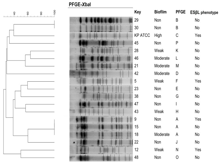
- DOI: 10.3390/pathogens10101345
Biofilm Formation of Clinical Klebsiella pneumoniae Strains Isolated from Tracheostomy Tubes and Their Association with Antimicrobial Resistance, Virulence and Genetic Diversity
Abstract
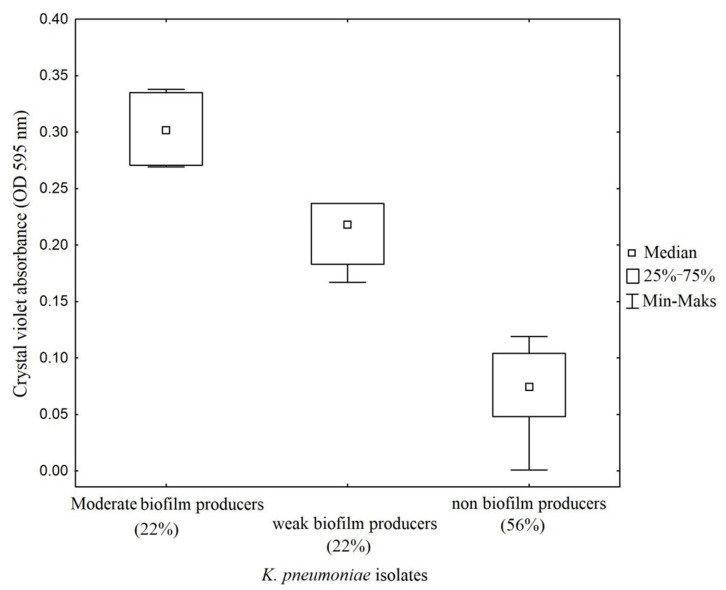
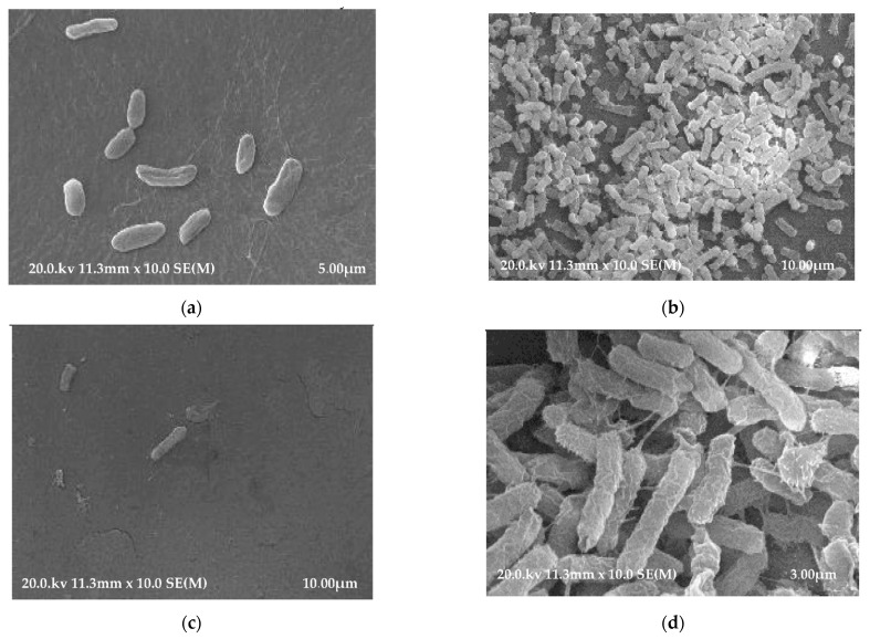
(1) Background: Due to the commonness of tracheotomy procedures and the wide use of biomaterials in the form of tracheostomy tubes (TTs), the problem of biomaterial-associated infections (BAIs) is growing. Bacterial colonization of TTs results in the development of biofilms on the surface of biomaterials, which may contribute to the development of invasive infections in tracheostomized patients. (2) Methods: Clinical strains of K. pneumoniae, isolated from TTs, were characterized according to their ability to form biofilms, as well as their resistance to antibiotics, whether they harbored ESβL genes, the presence of selected virulence factors and genetic diversity. (3) Results: From 53 patients, K. pneumoniae were detected in 18 of the TTs examined, which constituted 34% of all analyzed biomaterials. Three of the strains (11%) were ESβL producers and all had genes encoding CTX-M-1, SHV and TEM enzymes. 44.4% of isolates were biofilm formers, SEM demonstrating that K. pneumoniae formed differential biofilms on the surface of polyethylene (PE) and polyvinyl chloride (PVC) TTs in vitro. A large range of variation in the share of fimbrial genes was observed. PFGE revealed sixteen genetically distinct profiles. (4) Conclusions: Proven susceptibility of TT biomaterials to colonization by K. pneumoniae means that the attention of research groups should be focused on achieving a better understanding of the bacterial pathogens that form biofilms on the surfaces of TTs. In addition, research efforts should be directed at the development of new biomaterials or the modification of existing materials, in order to prevent bacterial adhesion to their surfaces.
Keywords: Klebsiella pneumoniae; PCR; PFGE; SEM; biofilm; tracheostomy tube.
Conflict of interest statement
The authors declare no conflict of interest.
Figures
Similar articles
-
Microbiological analysis of tracheostomy tube biofilms and antibiotic resistance profiles of potentially pathogenic microorganisms.Otolaryngol Pol. 2022 Jun 22;76(5):1-13. doi: 10.5604/01.3001.0015.8827. Otolaryngol Pol. 2022. PMID: 36622125
-
The Frequency of Occurrence of Resistance and Genes Involved in the Process of Adhesion and Accumulation of Biofilm in Staphylococcus aureus Strains Isolated from Tracheostomy Tubes.Microorganisms. 2022 Jun 14;10(6):1210. doi: 10.3390/microorganisms10061210. Microorganisms. 2022. PMID: 35744728 Free PMC article.
-
Distribution of bla(TEM), bla(SHV), bla(CTX-M) genes among clinical isolates of Klebsiella pneumoniae at Labbafinejad Hospital, Tehran, Iran.Microb Drug Resist. 2010 Mar;16(1):49-53. doi: 10.1089/mdr.2009.0096. Microb Drug Resist. 2010. PMID: 19961397
-
Promising strategies for future treatment of Klebsiella pneumoniae biofilms.Future Microbiol. 2020 Jan;15:63-79. doi: 10.2217/fmb-2019-0180. Epub 2020 Feb 12. Future Microbiol. 2020. PMID: 32048525 Review.
-
Identification and characterization of CTX-M-15 producing Klebsiella pneumoniae clone ST101 in a Hungarian university teaching hospital.Acta Microbiol Immunol Hung. 2015 Sep;62(3):233-45. doi: 10.1556/030.62.2015.3.2. Acta Microbiol Immunol Hung. 2015. PMID: 26551567 Review.
Cited by
-
A New Casjensviridae Bacteriophage Isolated from Hospital Sewage for Inactivation of Biofilms of Carbapenem Resistant Klebsiella pneumoniae Clinical Isolates.Pharmaceutics. 2024 Jul 5;16(7):904. doi: 10.3390/pharmaceutics16070904. Pharmaceutics. 2024. PMID: 39065601 Free PMC article.
-
Sonochemical Deposition of Gentamicin Nanoparticles at the PCV Tracheostomy Tube Surface Limiting Bacterial Biofilm Formation.Materials (Basel). 2023 May 16;16(10):3765. doi: 10.3390/ma16103765. Materials (Basel). 2023. PMID: 37241392 Free PMC article.
-
Relevance of the Adjuvant Effect between Cellular Homeostasis and Resistance to Antibiotics in Gram-Negative Bacteria with Pathogenic Capacity: A Study of Klebsiella pneumoniae.Antibiotics (Basel). 2024 May 26;13(6):490. doi: 10.3390/antibiotics13060490. Antibiotics (Basel). 2024. PMID: 38927157 Free PMC article. Review.
-
Emerging Issues on Antibiotic-Resistant Bacteria Colonizing Plastic Waste in Aquatic Ecosystems.Antibiotics (Basel). 2024 Apr 8;13(4):339. doi: 10.3390/antibiotics13040339. Antibiotics (Basel). 2024. PMID: 38667014 Free PMC article.
-
Molecular Profiling of a Multi-Strain Hypervirulent Klebsiella pneumoniae Infection Within a Single Patient.Infect Drug Resist. 2023 Mar 11;16:1367-1380. doi: 10.2147/IDR.S404202. eCollection 2023. Infect Drug Resist. 2023. PMID: 36937147 Free PMC article.
References
Grants and funding
LinkOut - more resources
Full Text Sources
