. 2021 Oct 9;26(20):6090.
doi: 10.3390/molecules26206090.

Glycyrrhizic Acid Inhibits SARS-CoV-2 Infection by Blocking Spike Protein-Mediated Cell Attachment

Affiliations

Glycyrrhizic Acid Inhibits SARS-CoV-2 Infection by Blocking Spike Protein-Mediated Cell Attachment

Jingjing Li et al. Molecules. .

Abstract

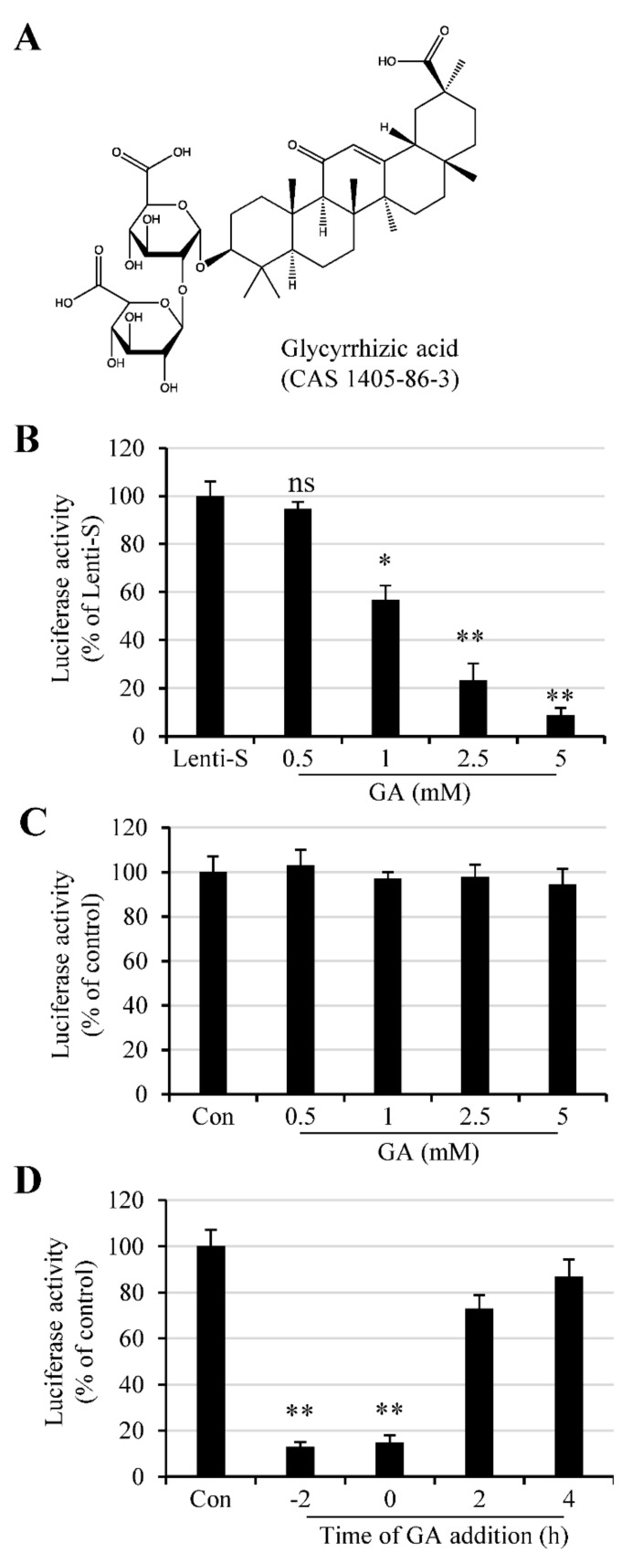
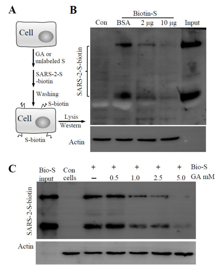
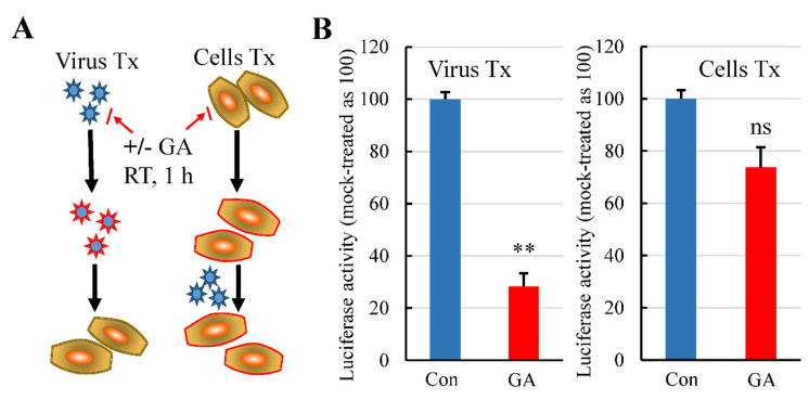
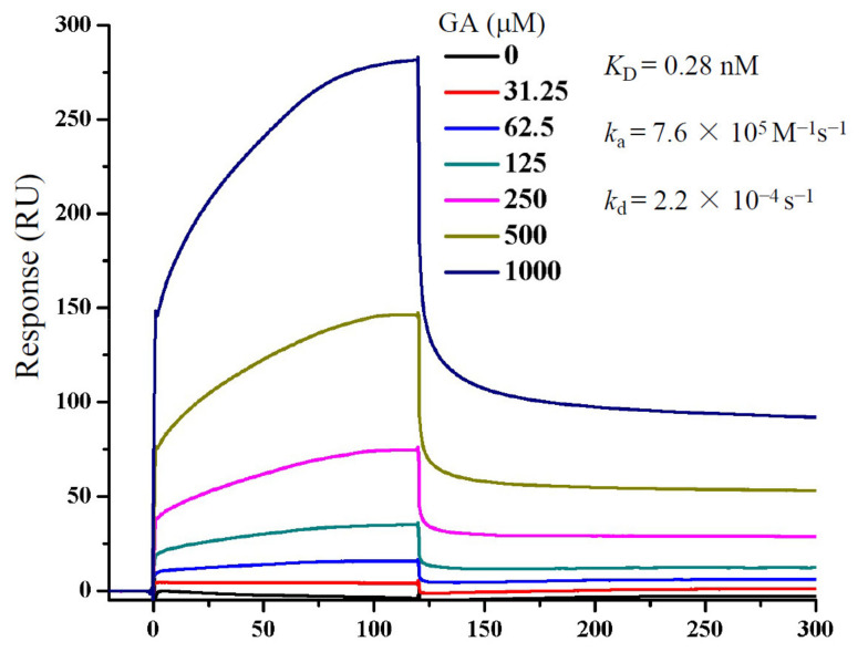
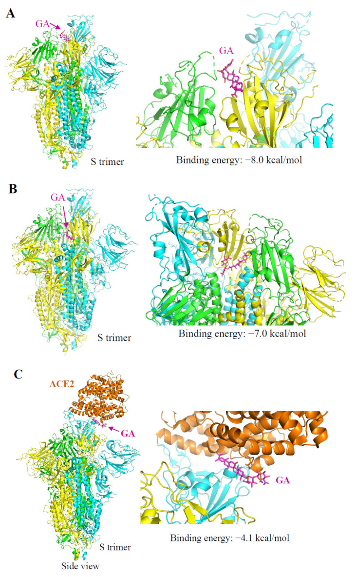
Glycyrrhizic acid (GA), also known as glycyrrhizin, is a triterpene glycoside isolated from plants of Glycyrrhiza species (licorice). GA possesses a wide range of pharmacological and antiviral activities against enveloped viruses including severe acute respiratory syndrome (SARS) virus. Since the S protein (S) mediates SARS coronavirus 2 (SARS-CoV-2) cell attachment and cell entry, we assayed the GA effect on SARS-CoV-2 infection using an S protein-pseudotyped lentivirus (Lenti-S). GA treatment dose-dependently blocked Lenti-S infection. We showed that incubation of Lenti-S virus, but not the host cells with GA prior to the infection, reduced Lenti-S infection, indicating that GA targeted the virus for infection. Surface plasmon resonance measurement showed that GA interacted with a recombinant S protein and blocked S protein binding to host cells. Autodocking analysis revealed that the S protein has several GA-binding pockets including one at the interaction interface to the receptor angiotensin-converting enzyme 2 (ACE2) and another at the inner side of the receptor-binding domain (RBD) which might impact the close-to-open conformation change of the S protein required for ACE2 interaction. In addition to identifying GA antiviral activity against SARS-CoV-2, the study linked GA antiviral activity to its effect on virus cell binding.

Keywords: SARS-CoV-2; autodocking; glycyrrhizin; surface plasmon resonance.


Conflict of interest statement

The authors declare no conflict of interest.

Figures

Figure 1
Figure 1
GA effect on Lenti-S infection. (A) Molecular structure of glycyrrhizic acid. (B) Effect of GA on Lenti-S infection. Vero E6 cells were infected with Lenti-S pseudovirus in the absence or presence of GA at indicated concentrations for 24 h. Luciferase activity was determined. Data are expressed as a percentage of untreated controls (Lenti-S). The experiment was performed twice, and data are mean ± SD of triplicate wells. Independent two-sample comparisons between non-GA treated sample and sample treated with GA at different concentrations, respectively, were determined by Student’s t test. ns: no significance; *, p < 0.05; **, p < 0.01. (C) Effect of GA on luciferase expression in pCMV-luc-transfected cells. Monolayers of 293T cells were transfected with pCMV-luc for 16 h. The cells were then treated with GA at indicated concentrations. Luciferase activity was determined after 24 h incubation. GA treatment did not affect luciferase expression delivered by an encoding plasmid. The experiment was performed twice. The readings from untreated samples were used as a control for the calculation of relative luciferase activity. Data are mean ± SD of duplicate wells from 2 independent experiments. (D) Time of GA addition on Lenti-S-mediated luciferase gene delivery. Vero E6 cells were untreated or treated with 3 mM GA at 2 h prior to (−2 h), during (0), or at 2 and 4 h post Lenti-S infection. Luciferase activity was determined 24 h PI. The experiment was performed 2 times. Data from the untreated controls were used for the calculation of relative luciferase activity. **, p < 0.01.
Figure 2
Figure 2
Effect of GA on recombinant S protein binding. (A) Schematic presentation of the experimental design. The cells were detached and incubated on ice for 60 min with GA or recombinant S protein (S unlabeled). Biotinylated S protein was then added for cell-attachment assay. After washing with ice-cold DMEM, cell-attached biotinylated S protein was detected by immunoblotting assay. (B) Biotinylated S protein binding to host cells in the presence of BSA or unlabeled S protein. Vero E6 cells resuspended in ice-cold DMEM containing 0.5% BSA were allowed to interact with Lenti-S in the presence or absence of unlabeled S protein (at 2 and 10 μg/mL). (C) Effect of GA on S protein binding to Vero E6 cells. A biotinylated S protein was allowed to bind to detached Vero E6 cells in the absence or presence of GA. Cell-bound biotinylated S protein was determined by immunoblotting assay. Actin was used as a loading control. Con, Vero E6 cells only. Input, biotinylated S protein for total binding.
Figure 3
Figure 3
Pretreatment of Lenti-S or host cells with GA on Lenti-S mediated luciferase gene delivery. (A) Diagram showing Lenti-S or host cells were treated (Tx) with 3 mM GA prior to the infection assay. Both Lenti-S and host cells were untreated or treated with 3 mM GA for 1 h. At the end, the GA-treated Lenti-S was 1:30 diluted and used to infect untreated host cells (final concentration of GA in the medium was approximately 0.1 mM). In parallel, the medium of the GA-treated cells was replaced with fresh medium without GA followed by infection of untreated Lenti-S. Luciferase activity was determined 24 h later. (B) GA effect on Lenti-S (Virus Tx) and on host cells (Cells Tx). Luciferase activity was expressed as a percentage of untreated controls. Data are mean ± SD of duplicate wells from 2 experiments. ns, no significance; **, p < 0.01 by Student’s t test.
Figure 4
Figure 4
Determination of GA binding to S protein by SPR. S protein was immobilized on to a CM5 sensor chip. GA at concentrations as indicated was passed over the chip, and SPR angle changes were recorded using the Biacore T200 system and reported as response units (RUs). Data fitting was performed using the 1:1 Langmuir model in the BIAevaluation software package (GE Healthcare).
Figure 5
Figure 5
Autodocking analysis of GA interaction with SARS-CoV-2 S and ACE2 protein. Three protomers of SARS-Cov-2 S protein (PDB id: 6vsb) are shown in cyan, green, and yellow, respectively. ACE2 protein (PDB id: 6m18) is in orange. The structure of proteins is presented in ribbons. The structure of GA (ZINC id:960251743495) is shown in magenta as sticks. Arrows indicate the predicted binding site of GA. (A) Predicted binding of GA on the S protein at the S–ACE2 interface with a calculated binding energy of –8.0 kcal/mol. (B) Predicted binding of GA on a binding pocket located at the inner side of the RBD with a binding energy of −7.0 kcal/mol. (C) Predicted binding of GA on the ACE2 protein at the ACE2–S interface with a binding energy of −4.1 kcal/mol.

References

    1. Li T., Lu H., Zhang W. Clinical observation and management of COVID-19 patients. Emerg. Microbes Infect. 2020;9:687–690. doi: 10.1080/22221751.2020.1741327. - DOI - PMC - PubMed
    1. Drayman N., DeMarco J.K., Jones K.A., Azizi S.A., Froggatt H.M., Tan K., Maltseva N.I., Chen S., Nicolaescu V., Dvorkin S., et al. Masitinib is a broad coronavirus 3CL inhibitor that blocks replication of SARS-CoV-2. Science. 2021;373:931–936. doi: 10.1126/science.abg5827. - DOI - PMC - PubMed
    1. National Health Commission of the People’s Republic of China, Diagnosis and Treatment Protocol for COVID-19 Patients (Tentative 8th Edition) [(accessed on 26 September 2021)]. Available online: http://regional.chinadaily.com.cn/pdf/DiagnosisandTreatmentProtocolforCO....
    1. Fiore C., Eisenhut M., Krausse R., Ragazzi E., Pellati D., Armanini D., Bielenberg J. Antiviral effects of Glycyrrhiza species. Phytother. Res. 2008;22:141–148. doi: 10.1002/ptr.2295. - DOI - PMC - PubMed
    1. Shibata S. A drug over the millennia: Pharmacognosy, chemistry, and pharmacology of licorice. Yakugaku Zasshi. 2000;120:849–862. doi: 10.1248/yakushi1947.120.10_849. - DOI - PubMed

MeSH terms

LinkOut - more resources