. 2021 Oct 16;26(20):6262.
doi: 10.3390/molecules26206262.

The Influence of Antitumor Unsymmetrical Bisacridines on 3D Cancer Spheroids Growth and Viability

Affiliations

The Influence of Antitumor Unsymmetrical Bisacridines on 3D Cancer Spheroids Growth and Viability

Jolanta Kulesza et al. Molecules. .

Abstract

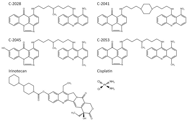
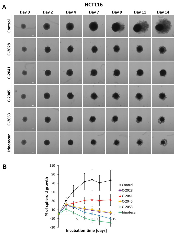
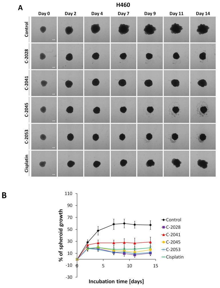
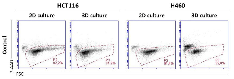
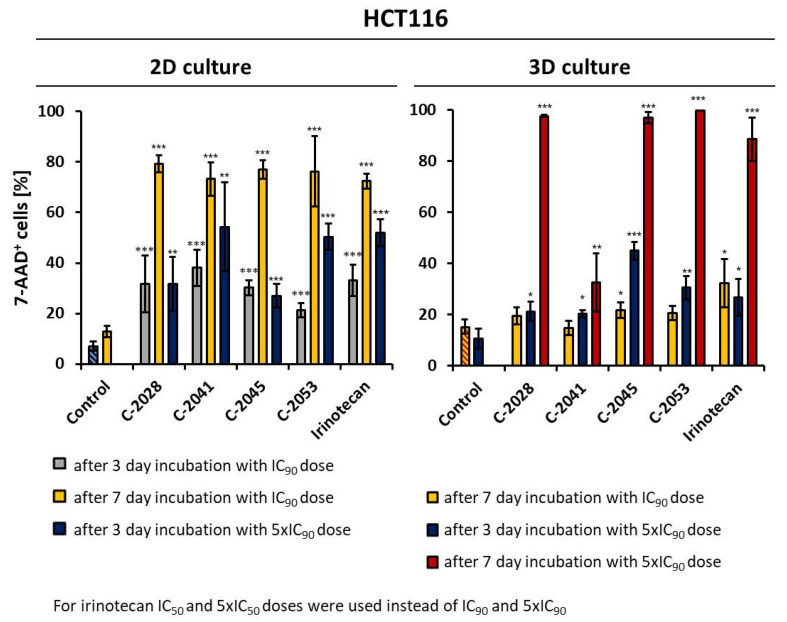
The culture of 3D spheroids is a promising tool in drug development and testing. Recently, we synthesized a new group of compounds, unsymmetrical bisacridines (UAs), which exhibit high cytotoxicity against various human cell lines and antitumor potency against several xenografts. Here, we describe the ability of four UAs-C-2028, C-2041, C-2045, and C-2053-to influence the growth of HCT116 and H460 spheres and the viability of HCT116 cells in 3D culture compared with that in 2D standard monolayer culture. Spheroids were generated using ultra-low-attachment plates. The morphology and diameters of the obtained spheroids and those treated with UAs were observed and measured under the microscope. The viability of cells exposed to UAs at different concentrations and for different incubation times in 2D and 3D cultures was assessed using 7-AAD staining. All UAs managed to significantly inhibit the growth of HCT116 and H460 spheroids. C-2045 and C-2053 caused the death of the largest population of HCT116 spheroid cells. Although C-2041 seemed to be the most effective in the 2D monolayer experiments, in 3D conditions, it turned out to be the weakest compound. The 3D spheroid culture seems to be a suitable method to examine the efficiency of new antitumor compounds, such as unsymmetrical bisacridines.

Keywords: 2D and 3D cultures; antitumor drugs; cancer treatment; cell death; spheroids; unsymmetrical bisacridines.


Conflict of interest statement

The authors declare no conflict of interest.

Figures

Figure 1
Figure 1
Chemical structures of the studied compounds: four unsymmetrical bisacridines (UAs), C-2028, C-2041, C-2045, and C-2053, and two reference compounds, irinotecan and cisplatin.
Figure 2
Figure 2
HCT116 spheroid morphology and kinetics. Spheroids were incubated with UAs at concentrations corresponding to IC90 values and irinotecan at an IC50 dose for 14 days. Every 2–3 days, images of the spheroids were taken and diameters were measured. (A) Representative images of the HCT116 control spheroids and spheroids treated with tested compounds. (B) Growth kinetics presented in a graph as percentages of spheroid growth over time. Data represent the averages of four independent experiments with standard deviation. White scale bars presented on images taken on day 0 correspond to 200 µm.
Figure 3
Figure 3
H460 spheroid morphology and kinetics. Spheroids were incubated with UAs at concentrations corresponding to IC90 values and cisplatin at an IC50 dose for 14 days. Every 2–3 days, images of the spheroids were taken and diameters were measured. (A) Representative images of the H460 control spheroids and spheroids treated with tested compounds. (B) Growth kinetics presented in a graph as percentages of spheroid growth over time. Data represent the averages of four independent experiments with standard deviation. White scale bars presented on images taken on day 0 correspond to 200 µm.
Figure 4
Figure 4
Viability of HCT116 (left) and H460 (right) cells cultured in 2D and 3D conditions. Cells in both culture systems were stained on day 3 with 7-AAD and subjected to flow cytometry analysis. P2: fraction of the 7-AAD negative cells (alive). Presented cytograms are representative of four independent experiments.
Figure 5
Figure 5
Effects of C-2028, C-2041, C-2045, C-2053, and irinotecan on cell viability in HCT116 cells cultured in 2D and 3D conditions. Cells were incubated with tested compounds at concentrations corresponding to IC90 and 5× IC90 values (IC50 and 5× IC50 for irinotecan) for 3 or 7 days, stained with 7-AAD, and subjected to flow cytometry analysis. Bar graphs show quantified data, expressed as the percentages of 7-AAD+ (dead) cells after incubation with the tested compounds in 2D (left) and 3D (right) cell cultures. Data are presented as the means ± SD of at least three independent experiments. Significant differences in cell percentages between the control and cells treated with the compound are indicated as follows: * p < 0.05; ** p < 0.01; *** p < 0.001.

References

    1. Melissa C., Siegel R. American Cancer Society: Global Cancer Facts & Figures. 4th ed. American Cancer Society; Atlanta, GA, USA: 2018.
    1. Breslin S., O’Driscoll L. Three dimensional cell culture: The missing link in drug discovery. Drug Discov. Today. 2013;18:240–249. doi: 10.1016/j.drudis.2012.10.003. - DOI - PubMed
    1. Edmondson R., Jenkins Brogile J., Adcock A.F., Yang L. Three-dimensional cell culture systems and their applications in drug discovery and cell-based biosensors. Assay Drug Dev. Technol. 2014;12:207–218. doi: 10.1089/adt.2014.573. - DOI - PMC - PubMed
    1. Waltz A., Unger C., Kramer N., Unterleuthner D., Scherzer M., Hengstschläger M., Schwanzer-Pfeiffer D., Dolznig H. The resazurin reduction assay can distinguish cytotoxic from cytostatic compounds in spheroid screening assays. J. Biomol. Screen. 2014;19:1047–1059. doi: 10.1177/1087057114532352. - DOI - PubMed
    1. Lee J., Lilly D., Doty C., Podsiadlo P., Kotov N.A. In vitro toxicity testing of nanoparticles in 3D cell culture. Small. 2009;5:1213–1221. doi: 10.1002/smll.200801788. - DOI - PubMed

MeSH terms

Substances