Case Reports
. 2021 Oct 11;11(10):1069.
doi: 10.3390/life11101069.

Cutaneous Localization of Classic Hodgkin Lymphoma Associated with Mycosis Fungoides: Report of a Rare Event and Review of the Literature

Affiliations
Case Reports

Cutaneous Localization of Classic Hodgkin Lymphoma Associated with Mycosis Fungoides: Report of a Rare Event and Review of the Literature

Magda Zanelli et al. Life (Basel). .

Abstract

Mycosis fungoides and nodal classic Hodgkin lymphoma (cHL) have been reported to occur concurrently or sequentially in the same patient. A long-lasting mycosis fungoides more often precedes the onset of nodal cHL, although few cases of nodal cHL followed by mycosis fungoides have been observed. Skin involvement is a rare manifestation of cHL that may be observed in the setting of advanced disease. The decrease in skin involvement in cHL is mainly due to the improved therapeutic strategies. The concurrent presence of mycosis fungoides and cutaneous localization of classic Hodgkin lymphoma represents a very uncommon event, with only two cases reported so far. Herein, we describe the case of a 71-year-old man, with a history of recurrent nodal cHL, who developed MF and, subsequently, the cutaneous localization of cHL. The clinicopathological features of the two diseases are described focusing on the main differential diagnoses to be taken into consideration, and a review of the literature is performed.

Keywords: classic Hodgkin lymphoma; mycosis fungoides; skin.


Conflict of interest statement

The authors declare no conflict of interest.

Figures
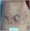
Figure 1
Figure 1
Cutaneous manifestation of cHL: a large, ulcerated nodule plus multiple smaller papules in the axillary region (original image from Dr. M.I.A.d.C).
Figure 2
Figure 2
(A) Histology of cutaneous localization of cHL: medium power view showing a polymorphic dermal infiltrate containing sparse, atypical, large-sized cells (yellow circles) (hematoxylin and eosin staining, 200× magnification; original image from Dr. M. Zanelli; (B) high-power view highlighting dispersed atypical cells with prominent nucleolus (yellow circle) within an inflammatory background rich in eosinophils (hematoxylin and eosin staining, 400× magnification; original image from Dr. M. Zanelli).
Figure 3
Figure 3
High-power view of CD30 immunostaining highlighting atypical cells with membranous and dot-like paranuclear positivity (yellow circle) (CD30 immunostaining, Ventana immunostainer, 400× magnification; original image from Dr. M. Zanelli).
Figure 4
Figure 4
The clinical features of early MF may be rather subtle: a slightly erythematous and finely scaling patch with a wrinkled appearance (original image from Prof. C.L).
Figure 5
Figure 5
Medium-power view of MF histology: the dermal infiltrate consists of small and medium-sized lymphocytes with evident epidermotropism (hematoxylin and eosin staining, 200×; original image from Dr. M. Zanelli).
Figure 6
Figure 6
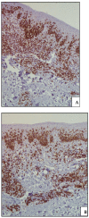
(A) Medium-power view of CD3 immunostaining confirming the T-cell nature of the dermal and epidermal lymphoid infiltrate (CD3 immunostaining, Ventana immunostainer, 200× magnification; original image from Dr. M. Zanelli); (B) CD8 positivity highlighting the cytotoxic phenotype of the dermal infiltrate with striking epidermotropism (CD8 immunostaining, Ventana immunostainer, 200× magnification; original image from Dr. M. Zanelli).

References

    1. WHO Classification of Tumours Editorial Board, editor. WHO Classification of Tumours Haematopoietic and Lymphoid Tissues. Revised 4th ed. IARC; Lyon, France: 2017.
    1. Parente P., Zanelli M., Sanguedolce F., Mastracci L., Graziano P. Hodgkin Reed-Sternberg-like cells in non-Hodgkin lymphoma. Diagnostics. 2020;10:1019. doi: 10.3390/diagnostics10121019. - DOI - PMC - PubMed
    1. Connors J.M. Clinical manifestations and natural history of Hodgkin lymphoma. Cancer J. 2009;15:124–128. doi: 10.1097/PPO.0b013e3181a282d8. - DOI - PubMed
    1. Zanelli M., Mengoli M.C., Valli R., Froio E., Bisagni A., Zizzo M., De Marco L., Ascani S. Primary classic Hodgkin lymphoma of the ileum and Epstein-Barr virus mucocutaneous ulcer of the colon: Two entities compared. Virchows Archiv. 2019;474:117–123. doi: 10.1007/s00428-018-2451-9. - DOI - PubMed
    1. Parente P., Zanelli M., Zizzo M., Carosi I., Di Candia L., Sperandeo M., Lacedonia D., Fesce V.F., Ascani S., Graziano P. Primary pulmonary Hodgkin lymphoma presenting as multiple cystic lung lesions: Diagnostic usefulness of cell block. Cytopathology. 2020;31:236–239. doi: 10.1111/cyt.12814. - DOI - PubMed

Publication types

LinkOut - more resources