. 2021 Sep 26;10(10):2547.
doi: 10.3390/cells10102547.

miR-21-3p/IL-22 Axes Are Major Drivers of Psoriasis Pathogenesis by Modulating Keratinocytes Proliferation-Survival Balance and Inflammatory Response

Affiliations

miR-21-3p/IL-22 Axes Are Major Drivers of Psoriasis Pathogenesis by Modulating Keratinocytes Proliferation-Survival Balance and Inflammatory Response

Florence Abdallah et al. Cells. .

Abstract

Psoriasis is a chronic inflammatory skin disease that is mediated by complex crosstalk between immune cells and keratinocytes (KCs). Emerging studies have showed a specific psoriatic microRNAs signature, in which miR-21 is one of the most upregulated and dynamic miRNAs. In this study, we focused our investigations on the passenger miR-21-3p strand, which is poorly studied in skin and in psoriasis pathogenesis. Here, we showed the upregulation of miR-21-3p in an IMQ-induced psoriasiform mouse model. This upregulation was correlated with IL-22 expression and functionality, both in vitro and in vivo, and it occurred via STAT3 and NF-κB signaling. We identified a network of differentially expressed genes involved in abnormal proliferation control and immune regulatory genes implicated in the molecular pathogenesis of psoriasis in response to miR-21-3p overexpression in KCs. These results were confirmed by functional assays that validated the proliferative potential of miR-21-3p. All these findings highlight the importance of miR-21-3p, an underestimated miRNA, in psoriasis and provide novel molecular targets for therapeutic purposes.

Keywords: IL-22; keratinocytes; miR-21-3p; miR-21-5p; proliferation; psoriasis.


Conflict of interest statement

The authors declare no competing interests.

Figures
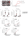
Figure 1
Figure 1
miR-21 (5p and 3p) expression patterns during psoriasis development. IMQ was applied daily for 6 consecutive days on the shaved backs of C57BL/6 wild-type (WT) and IL-22-knockout mice (IL-22−/−). Pathogenesis was studied from day 1 to day 7. (A) Pictures of mice representing skin lesions throughout the development of psoriasis skin lesions (left), cumulative score representations (a.u.) of erythema and scaling, on the back skin, scored daily on a scale from 0 (absence of severity) to 4 (highest severity) (right). (B,C) RT-qPCR quantification of the relative expression of miR-21-5p- or miR-21-3p-to-miR-16-5p levels in lesional skin of mice treated with IMQ. (D) Pearson correlations established in 30 mice comparing the relative expression of miR-21-5p and IL-22 relative expression or IL-22 concentration (pg/mL) from skin protein lysates, assayed by ELISA. (E) Pearson correlations established in 30 mice comparing the relative expression of miR-21-3p and IL-22 relative expression or IL-22 concentration (pg/mL) from skin protein lysates, assayed by ELISA. Data information: results are presented as mean values ± SEM. Data are representative of two independent experiments with five mice per group. The statistical comparisons between groups were performed using the two-tailed Student t-test: ** p < 0.01, *** p < 0.001.
Figure 2
Figure 2
IL-22 correlates with miR-21-3p and to a minor extent with miR-21-5p induction. (A) RT-qPCR quantification on the relative expression of miR-21-5p compared to miR-16-5p levels in HaCaT cells and NHEKs stimulated with 50 ng/mL IL-22 for 24 h. (B) RT-qPCR quantification of the relative expression of miR-21-3p compared to miR-16-5p levels in HaCaT cells and NHEKs stimulated with 50 ng/mL IL-22 for 24 h. (C) Pearson correlations between relative the expression of miR-21-5p and IL-22 in human skin biopsies from healthy (n = 10) and psoriatic individuals (n = 9). (D) Pearson correlations between the relative expression of miR-21-3p and IL-22 in human skin biopsies from healthy (n = 10) and psoriatic individuals (n = 9). Data information: results are presenteds as mean values ± SEM. Data are representative of three independent experiments done in triplicates. The statistical comparison between groups was performed by using a two-tailed Student’s t-test: * p < 0.05, ** p < 0.01.
Figure 3
Figure 3
miR-21 induction in psoriasis is IL-22-dependent. (A) Schematic representation of IMQ-induced psoriasiform or control mice (vehicle application)—time course of IL-22−/− mice that received 100 ng of IL-22 or an equivalent amount of physiological serum subcutaneously on the shaved area, one day before the induction of pathogenesis and up to day 7. (B) Relative expression of miR-21-5p to miR-16-5p at day 3 and day 6 post-IMQ application in vehicle mice (white histogram), IL-22-treated mice (light gray histogram), IMQ induced psoriasiform mice (dark gray histogram), and IMQ-induced psoriasiform mice treated with IL-22 (black histogram). (C) Relative expression of miR-21-3p to miR-16-5p at day 3 and day 6 post-IMQ application in vehicle mice (white histogram), IL-22-treated mice (light gray histogram), IMQ-induced psoriasiform mice (dark gray histogram), and IMQ-induced psoriasiform mice treated with IL-22 (black histogram). Data information: results are presented as mean values ± SEM. Data are representative of two independent experiments with at least five mice per group. The statistical comparison between groups was performed using two-way ANOVA: * p < 0.05, ** p < 0.01, *** p < 0.001.
Figure 4
Figure 4
Requirements of STAT3 and NF-κB signaling for the induction of miR-21-3p in IL-22-stimulated keratinocytes. (A) Schematic representation of the miR-21 promoter gene with the putative binding sites for STAT3 (S1, S2, and S3) and NF-κB (k1, k2, k3, and k4) transcription factors. (B) The luciferase activity of NF-κB in HaCaT cells transfected with NF-κB luciferase reporter construct was measured after 24 h of 50 ng/mL IL-22 stimulation. (C) IL-22 induction of the STAT3 pathway in HaCaT cells was evaluated using a cell-based ELISA kit. (D) Representation of the fold change in miR-21 (5p and 3p) relative expression, assessed via RT-qPCR. The fold change corresponds to the ratio of 50 ng/mL IL-22 over 0 ng/mL IL-22-treated HaCaT cells and silenced with si-Ctrl, si-p65, si-STAT3, or si-p65/STAT3 for 24 h. Data information: results are presented as mean values ± SEM. Data for B and C are representative of two independent experiments and for D are representative of three independent experiments performed in triplicate. The statistical comparison between groups was performed using the two-tailed Student’s t-test: * p < 0.05, ** p < 0.01, *** p < 0.001.
Figure 5
Figure 5
Functional assay using the RNAi-inducible luciferase expression system (RILES) to monitor miR-21 activity in HaCaT cells. (A) Schematic representation of the RILES construction. Briefly, the firefly luciferase reporter gene is repressed by a translational repressor (CymR—in red). The latter is under the control of the miR-21 cassette (miR-21.5pT or miR-21.3pT cassette, black histogram). Thus, the binding of miR-21-5p or miR-21-3p to the miR-21.5pT and miR-21.3pT cassette, respectively, results in CymR degradation that is translated by luciferase expression. Changes in luciferase levels reflect whether or not a miRNA can induce CymR mRNA degradation or translation arrest, which indicate miRNA functionality. An empty miR cassette, pRILES, was used as a control plasmid. (B,C) miR-21-5p and miR-21-3p expression induction assessments using RILES technology in HaCaT cells stimulated with 0, 20, 50, and 100 ng/mL of IL-22 for 24 h. Data are presented as relative luciferase units (RLU) normalized to the total amount of protein (mg). Data information: results are presented as mean values ± SEM. Data are representative of three independent experiments performed in triplicate. The statistical comparison between groups was performed by using two-tailed Student’s t-test: * p < 0.05.
Figure 6
Figure 6
DEG analysis in KCs overexpressing miR-21-3p versus negative control. (A,C) Functionally grouped networks for significant downregulated genes (blue) and upregulated genes (red). Only the labels of the 30 most significant terms per group are shown. Each node represents an enriched GO term. Related GO terms are connected by a line, the thickness of which reflects the percentage of overlapping genes. The size of the node corresponds to the number of genes. The network was automatically laid out using the ShiniGo tool with an edge cut-off of 0.2. Data information: data are representative of experimental triplicates per condition. (B) Venn diagram. showing the extent of overlap among all expressed genes between the miR-21-3p overexpressing HaCaT cells versus NCs. The level of significant gene modulation was p.adjusted < 0.05.
Figure 7
Figure 7
Comparative analysis between miR-21-3p-overexpressing KCs and psoriatic skin. (A) Graphs representing the numbers of significant DEGs (red: upregulated; blue: downregulated) between the indicated groups (PP vs. H, PN vs. H and PP vs. PN, from Pasquali et al., 2019 [9] and miR-21-3p, from this work). The Venn diagram shows the extent of overlap among DEGs. (B) Enrichment analysis of the 395 genes upregulated in PP keratinocytes and induced by miR-21-3p.
Figure 8
Figure 8
miR-21-3p gain of function promotes KC proliferation. (A) Crystal violet staining at different time pointa (24 h, 48 h, 72 h, and 96 h) of HaCaT cells transfected with negative control (NC) or miR-21-5p or miR-21-3p mimics. (B) XTT assays at different time points (24 h, 48 h, 72 h, and 96 h) of HaCaT cells transfected with negative controls (NC), miR-21-5p or miR-21-3p mimics. (C) Cell cycle analysis with double Ki-67/PI staining 24 h post-transfection of HaCaT cells with negative control (NC) or miR-21-5p or miR-21-3p mimics. (D) Ki67 immunostaining 24 h post transfection of HaCaT cells with negative control (NC) or miR-21-5p or miR-21-3p mimics. Data information: results are presented as mean values ± SEM. Data are representative of three independent experiments performed in triplicate. The statistical comparison between groups was performed by using the two-way ANOVA test: * p < 0.05, *** p < 0.001.

References

    1. Springate D.A., Parisi R., Kontopantelis E., Reeves D., Griffiths C.E.M., Ashcroft D.M. Incidence, prevalence and mortality of patients with psoriasis: A U.K. population-based cohort study. Br. J. Dermatol. 2017;176:650–658. doi: 10.1111/bjd.15021. - DOI - PMC - PubMed
    1. Griffiths C.E., Barker J.N. Pathogenesis and clinical features of psoriasis. Lancet. 2007;370:263–271. doi: 10.1016/S0140-6736(07)61128-3. - DOI - PubMed
    1. Wolk K., Haugen H.S., Xu W., Witte E., Waggie K., Anderson M., Vom Baur E., Witte K., Warszawska K., Philipp S., et al. IL-22 and IL-20 are key mediators of the epidermal alterations in psoriasis while IL-17 and IFN-γ are not. J. Mol. Med. 2009;87:523–536. doi: 10.1007/s00109-009-0457-0. - DOI - PubMed
    1. Eyerich S., Eyerich K., Pennino D., Carbone T., Nasorri F., Pallotta S., Cianfarani F., Odorisio T., Traidl-Hoffmann C., Behrendt H., et al. Th22 cells represent a distinct human T cell subset involved in epidermal immunity and remodeling. J. Clin. Investig. 2009;119:3573–3585. doi: 10.1172/JCI40202. - DOI - PMC - PubMed
    1. Wolk K., Witte E., Wallace E., Döcke W.D., Kunz S., Asadullah K., Volk H.D., Sterry W., Sabat R. IL-22 regulates the expression of genes responsible for antimicrobial defense, cellular differentiation, and mobility in keratinocytes: A potential role in psoriasis. Eur. J. Immunol. 2006;36:1309–1323. doi: 10.1002/eji.200535503. - DOI - PubMed

Publication types