Review
. 2021 Sep 27;10(10):2555.
doi: 10.3390/cells10102555.

Comparative Review of Microglia and Monocytes in CNS Phagocytosis

Affiliations
Review

Comparative Review of Microglia and Monocytes in CNS Phagocytosis

Megumi Andoh et al. Cells. .

Abstract

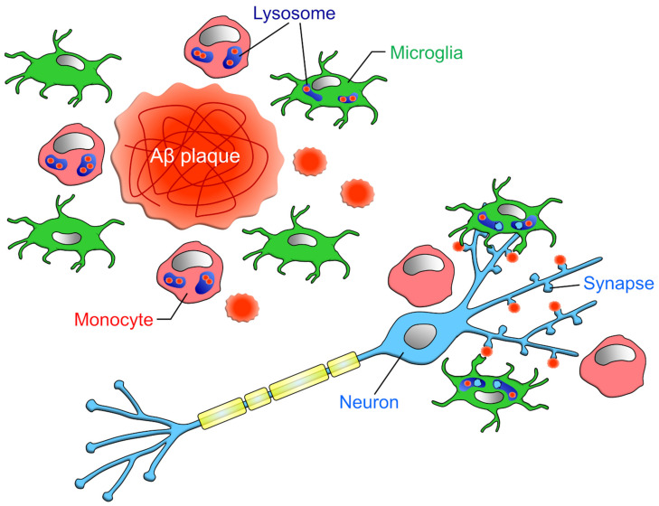
Macrophages maintain tissue homeostasis by phagocytosing and removing unwanted materials such as dead cells and cell debris. Microglia, the resident macrophages of the central nervous system (CNS), are no exception. In addition, a series of recent studies have shown that microglia phagocytose the neuronal synapses that form the basis of neural circuit function. This discovery has spurred many neuroscientists to study microglia. Importantly, in the CNS parenchyma, not only microglia but also blood-derived monocytes, which essentially differentiate into macrophages after infiltration, exert phagocytic ability, making the study of phagocytosis in the CNS even more interesting and complex. In particular, in the diseased brain, the phagocytosis of tissue-damaging substances, such as myelin debris in multiple sclerosis (MS), has been shown to be carried out by both microglia and blood-derived monocytes. However, it remains largely unclear why blood-derived monocytes need to invade the parenchyma, where microglia are already abundant, to assist in phagocytosis. We will also discuss whether this phagocytosis can affect the fate of the phagocytosing cell itself as well as the substance being phagocytosed and the surrounding environment in addition to future research directions. In this review, we will introduce recent studies to answer a question that often arises when studying microglial phagocytosis: under what circumstances and to what extent blood-derived monocytes infiltrate the CNS and contribute to phagocytosis. In addition, the readers will learn how recent studies have experimentally distinguished between microglia and infiltrating monocytes. Finally, we aim to contribute to the progress of phagocytosis research by discussing the effects of phagocytosis on phagocytic cells.

Keywords: infiltration; macrophage; microglia; monocyte; phagocytosis.


Conflict of interest statement

The authors declare no conflict of interest.

Figures

Figure 1
Figure 1
Development and expression signature of microglia and circulating monocytes: Erythro-myeloid progenitors (EMPs) from yolk sac invade into the CNS parenchyma during embryonic stage and differentiate into microglia. On the other hand, hematopoietic stem cells (HSCs) from fetal liver or bone marrow differentiate into monocytes and circulate in the blood vessels. In the physiological conditions, microglia are a main macrophage in the CNS parenchyma, but some diseases and injuries induce infiltration of circulating monocytes into the CNS parenchyma. We can distinguish between microglia and infiltrating monocytes by the genes listed in the figure. The molecules shown in green are called homeostatic microglial markers and are often considered as microglia specific. However, TMEM119, P2Y12, SIGLEC-H, and SALL1 are down-regulated in microglia in diseased brains [33,47,48], and peripheral macrophages begin to express molecules such as TMEM119, P2Y12, and SALL1 after infiltrating the brain parenchyma [49]. Therefore, it would be difficult to distinguish microglia from monocytes by these markers in situations where peripheral monocytes are infiltrating. On the other hand, HEXB has been suggested to be microglia-specific in both physiological and pathological conditions [50]. For signatures on other types of macrophages, please refer to the reviews by Prinz et al. and Li et al. [6,19].
Figure 2
Figure 2
Phagocytic capacities of microglia and infiltrating monocytes in the early phase of MS: Yamasaki et al. reported that at the onset of EAE, the ratio of microglia to monocytes contacting demyelinated axons is about 1:2, monocytes locating closer to axons than microglia [15]. The amount of myelin phagocytosis per cell is higher in microglia than monocytes [76,77], but monocytes as a whole (i.e., per animal) phagocytose more myelin.
Figure 3
Figure 3
Phagocytic capacities of microglia and infiltrating monocytes in ischemia: Rajan et al. reported that at 1 day after MCAO, when neuronal cell death is active, the majority of phagocytes in the parenchyma are microglia [42]. As more time passes, the infiltration of monocytes increases, and the ratio of microglia to monocyte becomes about 5:4.
Figure 4
Figure 4
Phagocytic capacities of microglia and infiltrating monocytes in AD: Unger et al. has reported that in the vicinity of Aβ plaque, the ratio of microglia to monocytes is about 1:1 [17]. However, when restricted to CD68-positive cells, the ratio of microglia to monocyte was about 1:3. Microglia phagocytose not only Aβ but also synapses attached to Aβ, while monocytes do not phagocytose synapses [34].
Figure 5
Figure 5
Significance of phagocytosis: Phagocytes possibly depend on the energy produced by the degradation of phagocytosed materials for their survival. It is also possible that phagocytes expose phagocytosed materials to the cell surface to mimic the target of phagocytosis. It has been suggested that phagocytes network or interact with other cells via presentation and exchange of phagocytosed materials.

References

    1. Galea I., Bechmann I., Perry V.H. What is immune privilege (not)? Trends Immunol. 2007;28:12–18. doi: 10.1016/j.it.2006.11.004. - DOI - PubMed
    1. Niederkorn J.Y. See no evil, hear no evil, do no evil: The lessons of immune privilege. Nat. Immunol. 2006;7:354–359. doi: 10.1038/ni1328. - DOI - PubMed
    1. Engelhardt B. Regulation of immune cell entry into the central nervous system. Results Probl. Cell Differ. 2006;43:259–280. doi: 10.1007/400_020. - DOI - PubMed
    1. Pösel C., Möller K., Boltze J., Wagner D.C., Weise G. Isolation and Flow Cytometric Analysis of Immune Cells from the Ischemic Mouse Brain. J. Vis. Exp. 2016;108:53658. doi: 10.3791/53658. - DOI - PMC - PubMed
    1. Korin B., Ben-Shaanan T.L., Schiller M., Dubovik T., Azulay-Debby H., Boshnak N.T., Koren T., Rolls A. High-dimensional, single-cell characterization of the brain’s immune compartment. Nat. Neurosci. 2017;20:1300–1309. doi: 10.1038/nn.4610. - DOI - PubMed

Publication types

LinkOut - more resources