Review
. 2021 Oct 1;10(10):2624.
doi: 10.3390/cells10102624.

Nuclear Dynamics and Chromatin Structure: Implications for Pancreatic Cancer

Affiliations
Review

Nuclear Dynamics and Chromatin Structure: Implications for Pancreatic Cancer

Luis F Flores et al. Cells. .

Abstract

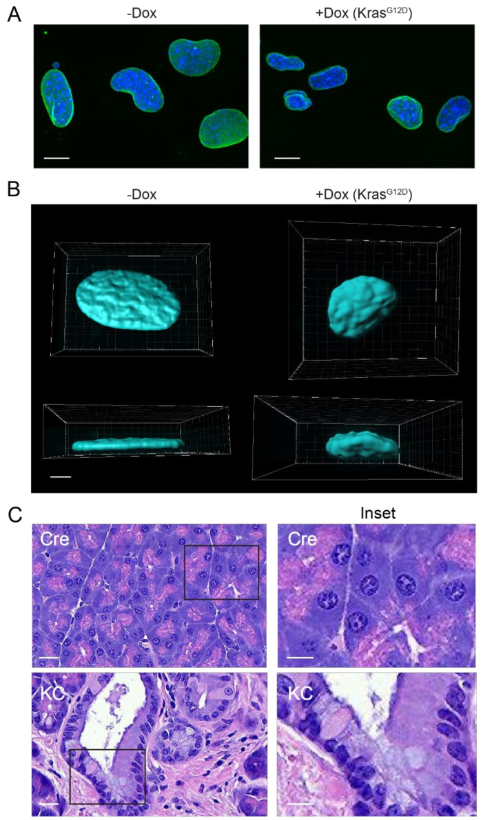
Changes in nuclear shape have been extensively associated with the dynamics and functionality of cancer cells. In most normal cells, nuclei have a regular ellipsoid shape and minimal variation in nuclear size; however, an irregular nuclear contour and abnormal nuclear size is often observed in cancer, including pancreatic cancer. Furthermore, alterations in nuclear morphology have become the 'gold standard' for tumor staging and grading. Beyond the utility of altered nuclear morphology as a diagnostic tool in cancer, the implications of altered nuclear structure for the biology and behavior of cancer cells are profound as changes in nuclear morphology could impact cellular responses to physical strain, adaptation during migration, chromatin organization, and gene expression. Here, we aim to highlight and discuss the factors that regulate nuclear dynamics and their implications for pancreatic cancer biology.

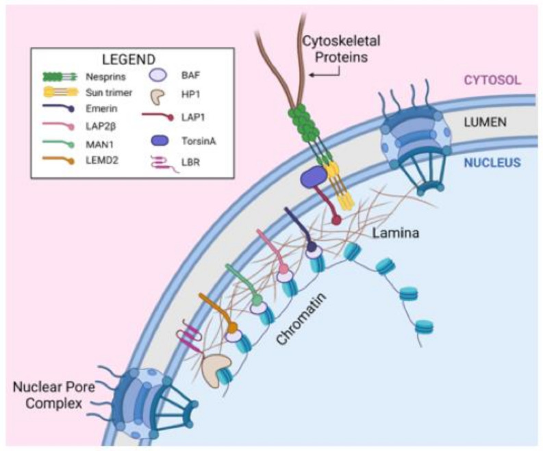
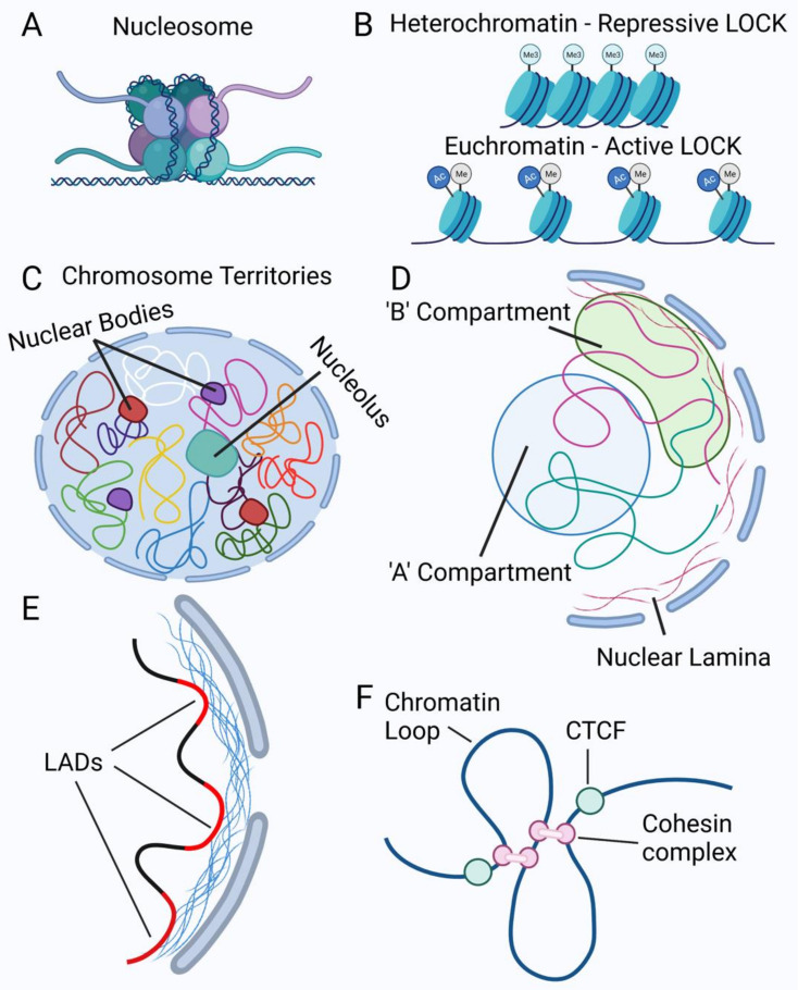
Keywords: chromatin; gene expression; nuclear lamina; nuclear morphology; pancreatic cancer.


Conflict of interest statement

The authors declare no conflict of interest.

Figures

Figure 1
Figure 1
Components of the nucleus and the nuclear envelope involved in nuclear dynamics. Nesprin proteins are a component of the outer nuclear membrane (ONM) that interact with various cytoskeletal proteins in the cytosol. SUN proteins are components of the inner nuclear membrane (INM) and can interact with nesprin proteins to form the Linker of Nucleoskeleton and Cytoskeleton (LINC) complex, which is a structural and mechanical feature of the nuclear envelope. TorsinA is contained in the nuclear envelope lumen and can interact with LINC complexes and the INM protein LAP1. Proteins from the LEM domain family (LAP2β, Emerin, MAN1, LEMD2) are INM proteins that interact with Barrier to Autointegration Factor (BAF) to facilitate heterochromatin anchoring to the lamina. The nuclear lamina is a meshwork of intermediate filaments (A- and B-type lamins) that sits inside the INM and gives structural support to the nucleus. Lamin B Receptor (LBR) is another INM protein that can interact with heterochromatin through the linker protein HP1. Nuclear Pore Complexes span both nuclear membranes to provide transport between the nucleus and cytosol. Created with biorender.com (accessed on 1 September 2021).
Figure 2
Figure 2
Chromatin organization in the nucleus. (A) Example of a nucleosome showing DNA wrapped around a histone octamer consisting of two H2A and H2B dimers, two H3 dimers, and two H4 dimers. (B) Example of heterochromatin or closed chromatin (top) and euchromatin or open chromatin (bottom). Long stretches of histones with similar lysine modifications illustrate Large Organized Chromatin Lysine (‘K’) modifications (LOCKs). The closed or open state refers to the accessibility of the chromatin to transcription factors or other DNA binding proteins. (C) Representation of chromosome territories where each colored line depicts one chromosome within the nucleus. Each chromosome is shown occupying its own space within the nucleus. The nucleolus is depicted here interacting with parts of some chromosomes. Nuclear bodies are also present, with examples of Cajal bodies (purple) and PML bodies (red) being shown. (D) Depiction of active ‘A’ compartment in the blue circle, indicating more open chromatin and actively transcribed genes, and ‘B’ compartment, which is mainly heterochromatin and therefore transcriptionally inactive. (E) Examples of Lamina-Associated Domains (LADs), shown here as red chromatin regions. (F) Examples of chromatin loops formed by cohesin complexes and demarcated by CTCF proteins. Created with biorender.com (accessed on 1 September 2021).
Figure 3
Figure 3
Oncogenic KRAS drives aberrant nuclear morphology in vitro and in vivo. (A) 4292F murine PDAC cells with a doxycycline (Dox)-inducible KRASG12D are shown, lamin B1 (green), DAPI (blue). Cells that are not expressing oncogenic KRAS (-Dox) present large and rather uniform nuclei. Induction of oncogenic KRAS with Dox causes a significant reduction in nuclear size and presents lamina alterations. Bar = 10 µm. (B) A 3D reconstruction from Z-stacks of DAPI signal from 4292F PDAC cells grown without or with Dox. All images were sized proportionally. (C) Murine H&E tissue sections from normal pancreas of Cre mice and PDAC pancreas from KRASG12D mice (KC). Normal pancreas displays larger and more uniform nuclei than those from PDAC. Bar = 100 µm. Inset regions are indicated by black rectangles and shown at higher magnification at right. Inset bar = 50 µm.

References

    1. Beale L.S. Results of the chemical and microscopical examination of solid organs and secretions. Examination of sputum from a case of cancer of the pharynx and the adjacent parts. Arch. Med. 1860;2:44–46.
    1. Papanicolaou G.N. A new procedure for staining vaginal smears. Science. 1942;95:438–439. doi: 10.1126/science.95.2469.438. - DOI - PubMed
    1. Papanicolaou G.N., Traut H.F. The diagnostic value of vaginal smears in carcinoma of the uterus 1941. Arch. Pathol. Lab. Med. 1997;121:211–224. - PubMed
    1. Dixon J.R., Selvaraj S., Yue F., Kim A., Li Y., Shen Y., Hu M., Liu J.S., Ren B. Topological domains in mammalian genomes identi-fied by analysis of chromatin interactions. Nature. 2012;485:376–380. doi: 10.1038/nature11082. - DOI - PMC - PubMed
    1. Guelen L., Pagie L., Brasset E., Meuleman W., Faza M.B., Talhout W., Eussen B.H., de Klein A., Wessels L., de Laat W., et al. Domain organization of human chromosomes revealed by mapping of nuclear lamina interactions. Nature. 2008;453:948–951. doi: 10.1038/nature06947. - DOI - PubMed

Publication types