. 2021 Oct 6;10(10):2671.
doi: 10.3390/cells10102671.

Increased Intrahepatic Expression of Immune Checkpoint Molecules in Autoimmune Liver Disease

Affiliations

Increased Intrahepatic Expression of Immune Checkpoint Molecules in Autoimmune Liver Disease

Zuzana Macek Jilkova et al. Cells. .

Abstract

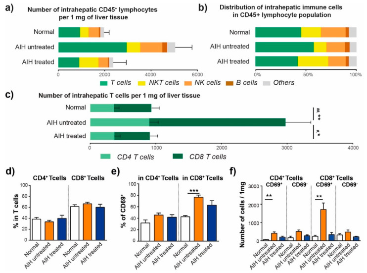
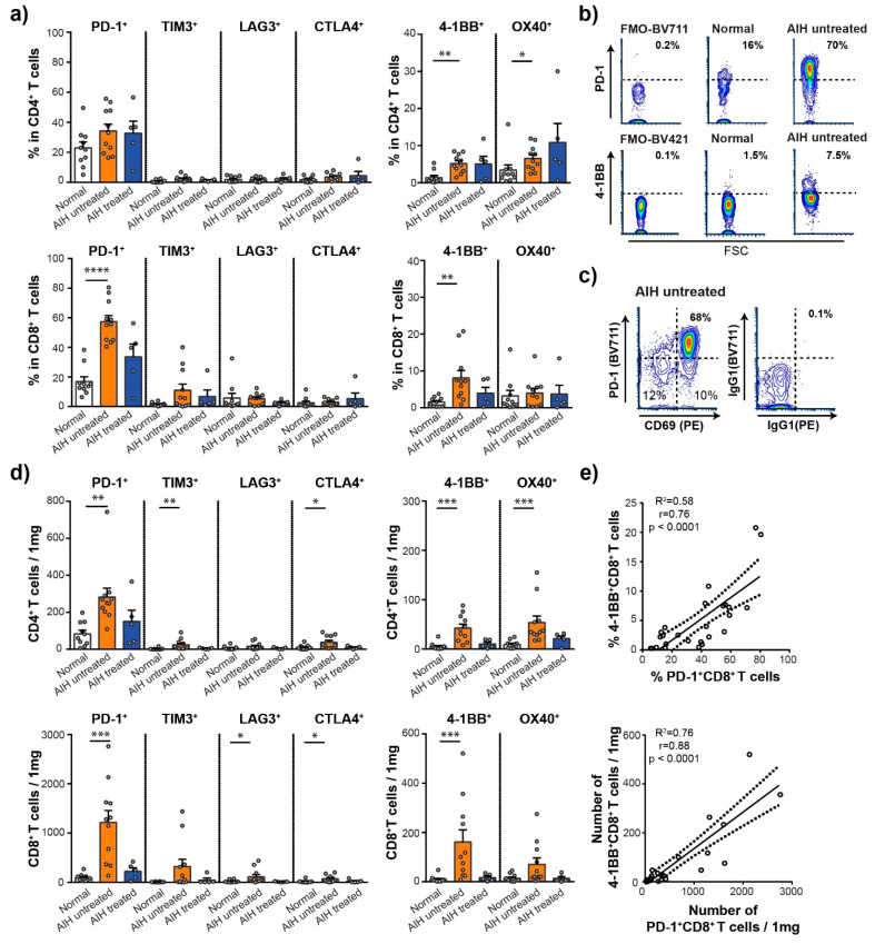
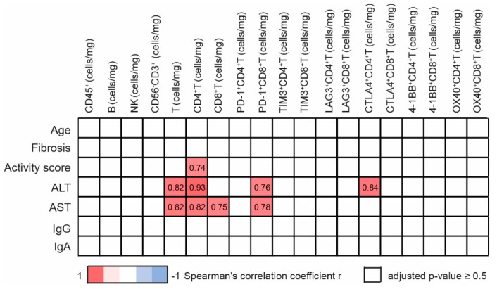
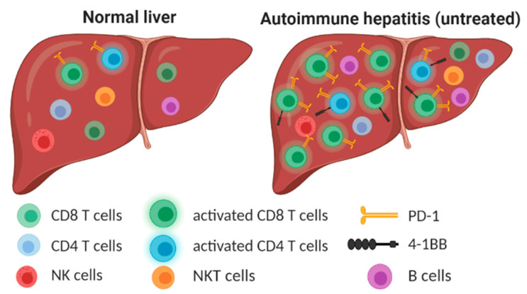
Immune checkpoint molecules (ICM) are critical in maintaining immunologic homeostasis and participate in preventing or promoting autoimmune disease development. Exploring a large panel of intrahepatic inhibitory and stimulatory ICM is necessary for drawing a general picture of the immune alterations in autoimmune hepatitis (AIH). Here, we performed a multiparametric analysis of ICM, including PD-1, TIM3, LAG3, CTLA-4, OX40 and 4-1BB, and we determined their expression on intrahepatic lymphocyte subsets in untreated and in treated patients with AIH in comparison to normal liver tissue. AIH patient-derived liver tissue revealed the overexpression of ICM, mainly PD-1 and 4-1BB, as well as the strong correlation between PD-1+ CD8+ T-cell abundance and severity of AIH (alanine transaminase and aspartate transaminase levels). Our results show that the ICM play an important role in the loss of immune homeostasis in the liver, providing an attractive approach to investigate their role as targets for effective therapeutic interventions.

Keywords: 4-1BB; PD-1; autoimmune hepatitis; autoimmune liver disease; immune checkpoint molecules.


Conflict of interest statement

The authors declare no conflict of interest.

Figures

Figure 1
Figure 1
Number and distribution of intrahepatic lymphocytes is modified in AIH. (a) Estimated number of CD45+ lymphocytes per 1 mg of liver tissue. (b) Frequency of intrahepatic immune cells in CD45+ lymphocytes. (c) Estimated number of intrahepatic T cells per 1 mg of liver tissue. # p < 0.05, ## p < 0.01 represent statistically significant difference in number of CD4+ T cells between groups, * p < 0.05, ** p < 0.01 represent statistically significant difference in number of CD8+ T cells between groups. Kruskal-Wallis test with Dunn multiple comparison post-test. (d) Frequency of intrahepatic CD4+ T cells and CD8+ T cells in T cell population. (e) Frequency of intrahepatic CD69+ cells in CD4+ T cell and in CD8+ T cell population. (f) Estimated number of CD69+ or CD69 CD4+ T cells and CD69+ or CD69 CD8+ T cells per 1 mg of liver tissue. * p < 0.05, ** p < 0.01, *** p < 0.001between groups, Kruskal-Wallis test with Dunn multiple comparison post-test. Data are expressed as mean ± SEM. Normal (n = 10), AIH untreated (n = 11) and AIH treated (n = 5).
Figure 2
Figure 2
Immune checkpoint molecule expression on intra-hepatic CD8 and CD4 T cells in different subgroups. (a) The frequency of intrahepatic immune checkpoint molecule positive CD4+ and CD8+ T cells. Normal (n = 10), AIH untreated (n = 11) and AIH treated (n = 5). Each circle represents a patient. Data are expressed as mean ± SEM, * p < 0.05, ** p < 0.01, **** p < 0.0001 between groups (Kruskal-Wallis test with Dunn multiple comparison post-test). (b) Representative flow cytometry contour plot showing the expression of intrahepatic PD-1 on CD8+ T cells and 4-1BB on CD4+ T cells in fluorescence minus one control sample (FMO), in normal liver biopsy and in biopsy from patient with untreated AIH. (c) Representative flow cytometry contour plot showing the co-expression of PD-1 and CD69 in intrahepatic CD8+ T cells of patient with untreated AIH. (d) An estimated number of intrahepatic immune checkpoint positive CD4+ T cells and CD8+ T cells per 1 mg of liver tissue. Normal (n = 10), AIH untreated (n = 11) and AIH treated (n = 5). Each circle represents a patient. Data are expressed as mean ± SEM, * p < 0.05, *** p < 0.001 between groups (Kruskal-Wallis test with Dunn multiple comparison post-test). (e) The correlation between the frequency of 4-1BB+ and PD-1+ CD8+ T cells (upper graph) and the correlation between the number of 4-1BB+ and PD-1+ CD8+ T cells (lower graph). (R2) R-squared, (r) Spearman correlation coefficient. Each circle represents a patient.
Figure 3
Figure 3
Correlation of patient characteristics and intrahepatic lymphocyte characteristics in untreated AIH group. Only significant correlations (Bonferroni-corrected p value < 0.5) are reported, numbers correspond to Spearman correlation coefficient r, positive correlation (red), negative correlation (blue), n = 11.
Figure 4
Figure 4
Untreated AIH is characterized by high accumulation of activated T cells with increased expression of PD-1 and 4-1BB immune checkpoint molecules. Created with BioRender.com.

References

    1. Carbone M., Neuberger J.M. Autoimmune liver disease, autoimmunity and liver transplantation. J. Hepatol. 2014;60:210–223. doi: 10.1016/j.jhep.2013.09.020. - DOI - PubMed
    1. Valgeirsson K.B., Hreinsson J.P., Björnsson E.S. Increased incidence of autoimmune hepatitis is associated with wider use of biological drugs. Liver Int. 2019;39:2341–2349. doi: 10.1111/liv.14224. - DOI - PubMed
    1. Takahashi A., Ohira H., Abe K., Zeniya M., Abe M., Arinaga-Hino T., Torimura T., Yoshizawa K., Takaki A., Kang J.-H., et al. Increasing incidence of acute autoimmune hepatitis: A nationwide survey in Japan. Sci. Rep. 2020;10:14250. doi: 10.1038/s41598-020-71296-0. - DOI - PMC - PubMed
    1. Grønbæk L., Otete H., Ban L., Crooks C., Card T., Jepsen P., West J. Incidence, prevalence and mortality of autoimmune hepatitis in England 1997-2015. A population-based cohort study. Liver Int. 2020;40:1634–1644. doi: 10.1111/liv.14480. - DOI - PubMed
    1. Mieli-Vergani G., Vergani D., Czaja A.J., Manns M.P., Krawitt E.L., Vierling J.M., Lohse A.W., Montano-Loza A.J. Autoimmune hepatitis. Nat. Reviews. Dis. Primers. 2018;4:18017. doi: 10.1038/nrdp.2018.17. - DOI - PubMed

Publication types