Review
. 2021 Oct 8;10(10):2687.
doi: 10.3390/cells10102687.

Clinical Applications of Cell-Scaffold Constructs for Bone Regeneration Therapy

Affiliations
Review

Clinical Applications of Cell-Scaffold Constructs for Bone Regeneration Therapy

Venkata Suresh Venkataiah et al. Cells. .

Abstract

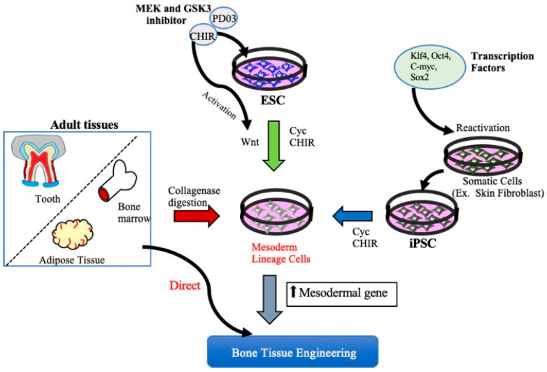
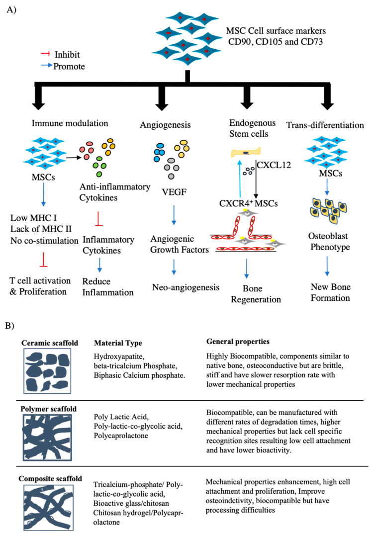
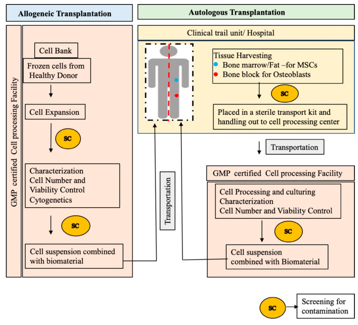
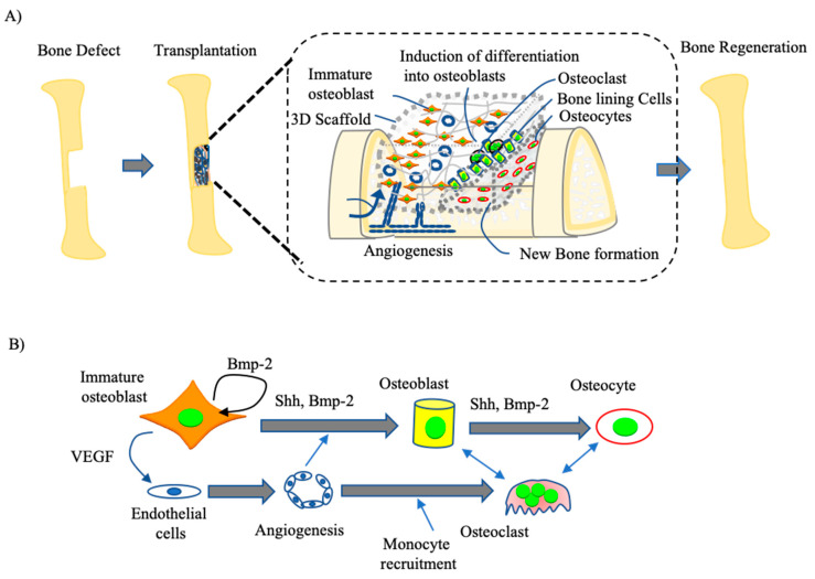
Bone tissue engineering (BTE) is a process of combining live osteoblast progenitors with a biocompatible scaffold to produce a biological substitute that can integrate into host bone tissue and recover its function. Mesenchymal stem cells (MSCs) are the most researched post-natal stem cells because they have self-renewal properties and a multi-differentiation capacity that can give rise to various cell lineages, including osteoblasts. BTE technology utilizes a combination of MSCs and biodegradable scaffold material, which provides a suitable environment for functional bone recovery and has been developed as a therapeutic approach to bone regeneration. Although prior clinical trials of BTE approaches have shown promising results, the regeneration of large bone defects is still an unmet medical need in patients that have suffered a significant loss of bone function. In this present review, we discuss the osteogenic potential of MSCs in bone tissue engineering and propose the use of immature osteoblasts, which can differentiate into osteoblasts upon transplantation, as an alternative cell source for regeneration in large bone defects.

Keywords: MSCs; bone tissue engineering; osteoblasts; scaffolds.


Conflict of interest statement

The authors declare no conflict of interest.

Figures

Figure 1
Figure 1
Schematic diagram of different approaches to obtain Mesenchymal stem cells. MSCs can be derived from either iPSCs, ESCs, or adult mesenchymal tissue. MSCs can be obtained by ESCs and iPSCs using small molecules such as mitogen-activated protein kinase (MEK) inhibitor, (MEK) inhibitor, PD0325901, glycogen synthase kinase 3 (GSK3) inhibitor, and CHIR99021 (CHIR). MSCs are also be derived from various connective tissues such as bone marrow, adipose tissue, and dental tissues by collagenase digestion or aspirates from bone marrow and adipose tissue directly used for BTE therapeutics. KLf4: Kruppel Like Factor 4, Oct4: Octamer-binding transcription factor 4, C-myc: Cellular-Myelocytomatosis, Sox-2: sex-determining region Y-box 2.
Figure 2
Figure 2
The combination of MSCs and bioscaffold materials used for BTE. (A) Mechanisms underlying MSC-based bone regeneration. Due to their characteristic expression of cell markers CD90, CD105, and CD73, and lack of HLA-molecules, MSCs have a bone tissue regeneration capacity through the actions of several mechanisms, including (1) the modulation of immune responses through the prevention of T-cell activation and reduction in the secretion of inflammatory cytokines; (2) the secretion of the angiogenic induction factor VEGF, which helps to form new blood vessels and in turn enhance bone regeneration; (3) the release of chemotactic chemokines at the bone defect site to recruit endogenous stem cells that will further enhance bone regeneration at that location; (4) the trans-differentiation of these cells into osteoblasts under the influence of host-derived factors that helps to promote new bone formation. (B) Representation of the routinely used scaffolds with examples and their general properties in the development of BTE technology.
Figure 3
Figure 3
Schematic outline of standard procedure in preparing cell-scaffold constructs for bone regeneration.
Figure 4
Figure 4
Schematics showing mechanisms of bone regeneration by immature osteoblasts. (A) Structures of the osteoblasts seeded scaffold constructs in the bone defect area following transplantation and healing of the defect site. (B) Molecular mechanism of bone remodeling by immature osteoblast. The immature osteoblasts under the influence of various cytokines such BMP2, SHH secreted from the bone matrix differentiate into osteoblasts. These osteoblasts produce various cell products, including enzymes alkaline phosphatase and collagenase, growth factors, osteocalcin, and collagen, part of the organic unmineralized component of bone. Few osteoblasts embed inside matrix to become osteocyte and others remain as a bone lining cells on the outer surface. Consequently, when osteoblasts lay down new matrix the osteoclast will differentiate from circulating monocytes/macrophages induced from osteoblasts secreted cytokines such as RANKL and M-CSF, as an inflammatory response to the bone defect from Osteoblasts. Simultaneously, angiogenic factors including VEGF are released from the osteoblasts to form new blood vessels.

References

    1. Nauth A., Schemitsch E., Norris B., Nollin Z., Watson J.T. Critical-Size Bone Defects: Is There a Consensus for Diagnosis and Treatment? J. Orthop. Trauma. 2018;32:S7–S11. doi: 10.1097/BOT.0000000000001115. - DOI - PubMed
    1. Roddy E., DeBaun M.R., Daoud-Gray A., Yang Y.P., Gardner M.J. Treatment of critical-sized bone defects: Clinical and tissue engineering perspectives. Eur. J. Orthop. Surg. Traumatol. 2018;28:351–362. doi: 10.1007/s00590-017-2063-0. - DOI - PubMed
    1. Yuan H., Xing K., Hsu H.-Y. Trinity of Three-Dimensional (3D) Scaffold, Vibration, and 3D Printing on Cell Culture Application: A Systematic Review and Indicating Future Direction. Bioengineering. 2018;5:57. doi: 10.3390/bioengineering5030057. - DOI - PMC - PubMed
    1. Plotkin L.I., Bellido T. Osteocytic signalling pathways as therapeutic targets for bone fragility. Nat. Rev. Endocrinol. 2016;12:593–605. doi: 10.1038/nrendo.2016.71. - DOI - PMC - PubMed
    1. Wang T., Yang X., Qi X., Jiang C. Osteoinduction and proliferation of bone-marrow stromal cells in three-dimensional poly (ε-caprolactone)/ hydroxyapatite/collagen scaffolds. J. Transl. Med. 2015;13:152. doi: 10.1186/s12967-015-0499-8. - DOI - PMC - PubMed

LinkOut - more resources