. 2021 Oct 13;10(10):2732.
doi: 10.3390/cells10102732.

Cullin4 E3 Ubiquitin Ligases Regulate Male Gonocyte Migration, Proliferation and Blood-Testis Barrier Homeostasis

Affiliations

Cullin4 E3 Ubiquitin Ligases Regulate Male Gonocyte Migration, Proliferation and Blood-Testis Barrier Homeostasis

Yan Yin et al. Cells. .

Abstract

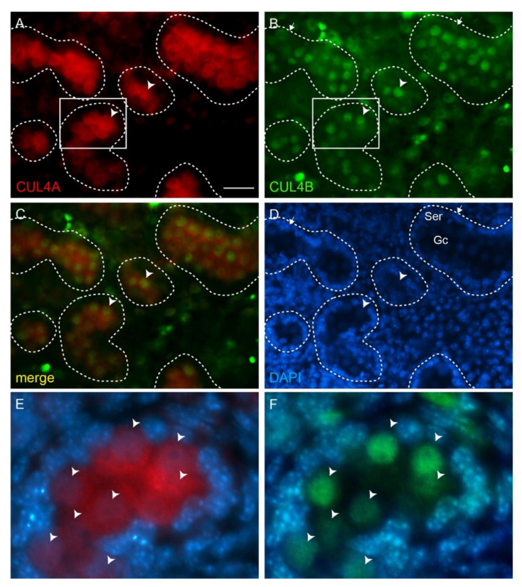
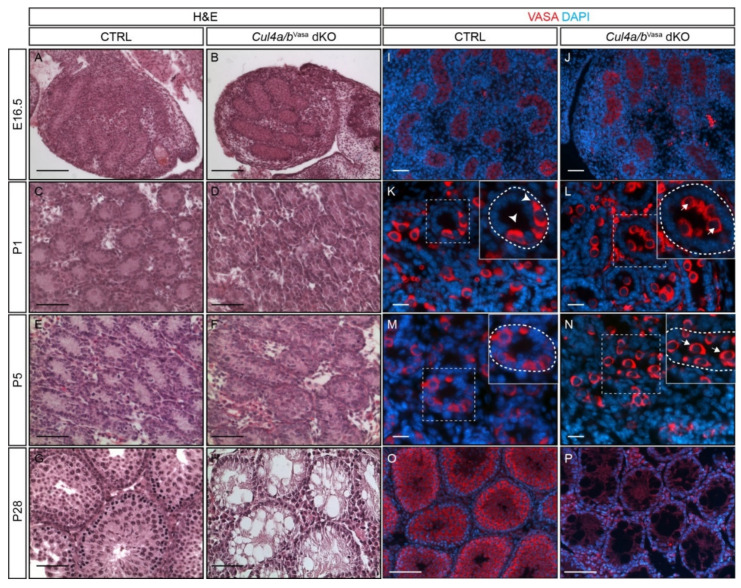
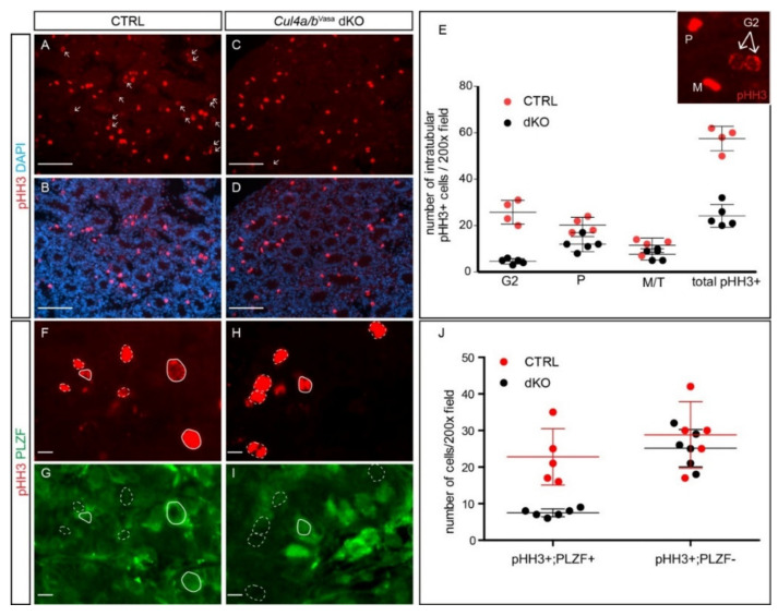
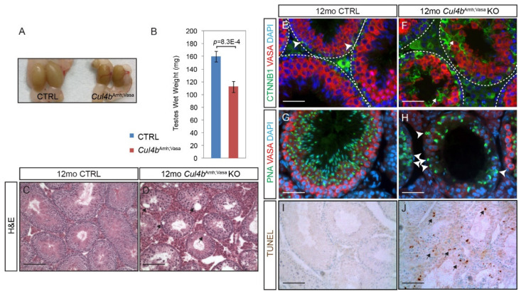
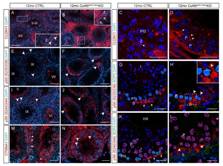
Ubiquitination, an essential posttranslational modification, plays fundamental roles during mammalian spermatogenesis. We previously reported the requirement of two Cullin 4 ubiquitin ligase family genes, Cullin 4a (Cul4a) and Cullin 4b (Cul4b), in murine spermatogenesis. Both genes are required for male fertility despite their distinct functions in different cell populations. Cul4a is required in primary spermatocytes to promote meiosis while Cul4b is required in secondary spermatocytes for spermiogenesis. As the two genes encode proteins that are highly homologous and have overlapping expression in embryonic germ cells, they may compensate for each other during germ cell development. In the present study, we directly address the potential functional redundancy of these two proteins by deleting both Cul4 genes, specifically, in the germ cell lineage during embryonic development, using the germ-cell specific Vasa-Cre line. Conditional double-knockout (dKO) males showed delayed homing and impaired proliferation of gonocytes, and a complete loss of germ cells before the end of the first wave of spermatogenesis. The dKO male germ cell phenotype is much more severe than those observed in either single KO mutant, demonstrating the functional redundancy between the two CUL4 proteins. The dKO mutant also exhibited atypical tight junction structures, suggesting the potential involvement of CUL4 proteins in spermatogonial stem cell (SSC) niche formation and blood-testis-barrier (BTB) maintenance. We also show that deleting Cul4b in both germ and Sertoli cells is sufficient to recapitulate part of this phenotype, causing spermatogenesis defects and drastically reduced number of mature sperms, accompanied by defective tight junctions in the mutant testes. These results indicate the involvement of CUL4B in maintaining BTB integrity.

Keywords: Cullin4; blood-testis barrier; spermatogenesis; ubiquitination.


Conflict of interest statement

The authors declare no conflict of interest.

Figures

Figure 1
Figure 1
CUL4A and CUL4B co-localize in fetal gonocytes. (AF) Immunofluorescence (IF) staining of CUL4A (red) and CUL4B (green) in E16.5 wild-type testis. Both CUL4A (A) and CUL4B (B) are highly expressed in the gonocytes (arrowheads), with CUL4B more prominent in the nuclei and CUL4A in both nuclei and cytoplasm. (C) shows merged CUL4A and CUL4B staining, and (D) shows DAPI counterstain. (E,F) are higher-magnitude views of the boxed areas in (A,B), respectively, with DAPI staining overlaid. Arrowheads point to gonocytes. Dashed lines outline seminiferous tubules. Ser, Sertoli cells; Gc, gonocytes. Scale bar 50 µm.
Figure 2
Figure 2
Cul4 genes are essential for spermatogenic cell survival and germ cell homing. (AH) Morphology of testes from E16.5, P1, P5 and P28 CTRL and Cul4a/bVasa dKO mice by H and E staining. Relative normal morphology was observed in neonatal dKO mutants, however, by P28 the mutant testes are filled with empty tubules. (IP) IF staining of germ cell marker, VASA, in testicular sections of E16.5, P1, P5 and P28 CTRL and Cul4a/bVasa dKO mice. In neonate mutants, VASA-positive germ cells are present in the dKO seminiferous tubules, but show delay in homing. Insets in (KN) show magnified view of boxed areas; dashed lines outline individual tubules. Note that clusters of germ cells are positioned in the mutant seminiferous tubule lumens (arrows, insets in L,N), whereas in the CTRLs they have migrated to the basement membranes (arrowheads, insets in K,M). By P28, VASA-positive germ cells are no longer detectable in dKO testes. Scale bars: 100 µm in (AD), (O,P); 50 µm in (EN).
Figure 3
Figure 3
Cul4 genes are critical to maintain male germ cell proliferation. (AD) IF staining of phospho-histone H3 (pHH3, red) in P5 CTRL and Cul4a/bVasa dKO testes. Arrows point to pHH3-positive G2 phase cells. (E) Quantification of pHH3-positive cells in seminiferous tubules revealed significant reduction in number of pHH3+ cells in the dKO, particularly in cells at G2 phase. Inset shows typical pHH3 staining pattern of cells at prophase (P), metaphase (M) and G2 phase. M/T, metaphase/telophase. Total: CTRL 57.5 ± 5.3, dKO 24.0 ± 5.3, p = 2.5 × 10 −5; G2: CTRL 25.8 ± 5.1, dKO 4.8 ± 1.3, p = 3.9 × 10 −5; P: CTRL 20.2 ± 3.3, dKO 13.0 ± 2.7, p = 0.007; M/T: CTRL 11.5 ± 3.1, dKO 13.0 ± 2.7, p = 0.07; n = 4 for CTRL and n = 5 for dKO. (FI) Double IF staining of pHH3 (red) and PLZF (green) of P5 CTRL and dKO testicular sections. Solid white lines circle out pHH3+; PLZF+ proliferating germ cells, dashed white lines circle out pHH3+; PLZF- cells. (J) Quantification of pHH3/PLZF double IF cells revealed significant decrease in number of double positive cells. pHH3+; PLZF+: CTRL 22.8 ± 7.7, dKO 7.5 ± 1.0, p = 8.8 × 10 −4; pHH3+; PLZF−: CTRL 28.8 ± 9.1, dKO 25.1 ± 5.1, p = 0.42; n = 5 for CTRL and n = 6 for dKO. Scale bars: 50 µm in (AD), 20 µm (FI).
Figure 4
Figure 4
Cul4 genes are required for male germ cell survival. (AD) IF staining of CUL4A (red) and SCP3 (green) of P28 testis. While primary spermatocytes expressing both CUL4A (A) and SCP3 (B) are present in the CTRL testis, this population is completely missing in the dKO mutant testis (C,D). (EH) IF staining of CUL4B (red) and SCP3 (green) of P28 testis. CUL4B is highly expressed in the Sertoli cells (E, arrowheads) and round spermatids (Spd) of CTRL testis (E,F); whereas in the Cul4a/bVasa dKO, CUL4B-positive Sertoli cells are the only cell type remaining in the seminiferous tubules (G,H arrowheads). (IN) IF staining of AR (red) and CTNNB1 (green) of P28 testicular sections. Wild-type testis show AR-high myoid cells (I, arrows) and AR-low Sertoli cells (I, arrowheads), and well organized germ cell/Sertoli cell network outlined by CTNNB1 staining (J). The dKO mutant testis retains myoid and Sertoli cells (L, arrows and arrowheads, respectively), with disorganized residual CTNNB1 staining (M). DAPI counterstain is shown in (K) and (N). Spd, spermatids; Spct, spermatocytes; Spg, spermatogonia; Int, interstitial cells. (O,P) IF staining of CLDN11 (red) and SCP3 (green) of P28 testis. At this stage in the wildtype testis, tight junctions have formed apical to the spermatogonial stem cells to separate the basal and adluminal compartments (O, arrowheads). CLDN11 was only detected in the basal-lateral membranes of dKO Sertoli cells (P, arrows). Scale bars: 50 µm.
Figure 5
Figure 5
Ablation of Cul4b in both germ cells and Sertoli cells causes extensive germ cell death. (A) Representative gross morphology of Cul4bAmh;Vasa KO and CTRL testes at 12 months. (B) Wet weight of testes isolated from 12mo mice. Data presented as mean ± s.d, n = 3 biologically independent testes. (C,D) Morphology of 12mo testicular sections by H and E staining. Mutant seminiferous tubule diameters are in general smaller, and many voids are visible inside the tubules indicating defective epithelial structure (arrows). (E,F) IF staining of VASA (red) and β-catenin (green). VASA-positive germ cells are orderly arranged, and β-catenin is most strongly expressed surrounding the basal-localized spermatogonia in the CTRL testis (E, arrowheads). Mutant germ cells are disorganized inside the tubules, and intensive β-catenin staining is detected throughout the length of Sertoli cells (F, arrows). (G,H) IF staining of VASA (red) and lectin PNA (green). PNA-positive spermatids are close to the lumen and positioned inside of the ring of VASA-strong primary spermatocytes, as spermatogenesis progresses in the CTRL testis. In the mutant, PNA-positive spermatids are substantially reduced in number, and many are abnormally positioned next to the basement membrane (H, arrowheads). (I,J) TUNEL-staining revealed extensive cell death inside the mutant seminiferous tubules (arrows). Scale bars: 200 µm in (C,D), 50 µm in (EH), 100 µm in (IJ).
Figure 6
Figure 6
Ablation of Cul4b in both germ cells and Sertoli cells leads to BTB defects. (A,B) IF staining and (C,D) confocal microscopy of tight junction marker CLDN11 in CTRL and Cul4bAmh;Vasa testis. The insets in A and B are magnified views of boxed areas. Basement membrane outlined by dashed lines in insets. (E,F) IF staining of pS6 (S235/236) showing its accumulation in the mutant tubules. (G,H) Confocal IF of pS6 (S235/236) (red) and SCP3 (green) showing localization of pS6 (S235/236) in CTRL spermatogonia (G, arrows), and ectopic activation in the mutant Sertoli cells (H, arrowheads). (I,J) IF staining of pS6 (S240/244) showing its accumulation in the mutant tubules. (K,L) Confocal IF of pS6(S240/244) (red) and SCP3 (green) showing its normal expression in spermatogonia (K, arrows) and ectopic activation in the mutant germ cells (L, arrowheads). (M,N) IF of α-catenin (CTNNA1) showing its accumulation in the mutant testis. S, spermatogonia; P, pachytene spermatocytes; Z, zygotene spermatocytes; Spg,. White dashed lines outline the seminiferous tubules. Scale bars: 200 µm in (A,B), (E,F), (I,J); 50 µm in the rest.

Similar articles

Cited by

References

    1. Schiff J.D., Ramírez M.L., Bar-Chama N. Medical and surgical management male infertility. Endocrinol. Metab. Clin. N. Am. 2007;36:313–331. doi: 10.1016/j.ecl.2007.03.003. - DOI - PubMed
    1. Anderson R., Copeland T.K., Schöler H., Heasman J., Wylie C. The onset of germ cell migration in the mouse embryo. Mech. Dev. 2000;91:61–68. doi: 10.1016/S0925-4773(99)00271-3. - DOI - PubMed
    1. Runyan C., Schaible K., Molyneaux K., Wang Z., Levin L., Wylie C. Steel factor controls midline cell death of primordial germ cells and is essential for their normal proliferation and migration. Development. 2006;133:4861–4869. doi: 10.1242/dev.02688. - DOI - PubMed
    1. Moreno S.G., Attali M., Allemand I., Messiaen S., Fouchet P., Coffigny H., Romeo P.H., Habert R. TGFbeta signaling in male germ cells regulates gonocyte quiescence and fertility in mice. Dev. Biol. 2010;342:74–84. doi: 10.1016/j.ydbio.2010.03.007. - DOI - PubMed
    1. Nagano R., Tabata S., Nakanishi Y., Ohsako S., Kurohmaru M., Hayashi Y. Reproliferation and relocation of mouse male germ cells (gonocytes) during prespermatogenesis. Anat. Rec. 2000;258:210–220. doi: 10.1002/(SICI)1097-0185(20000201)258:2<210::AID-AR10>3.0.CO;2-X. - DOI - PubMed

Publication types