

Role of Aldynoglia Cells in Neuroinflammatory and Neuroimmune Responses after Spinal Cord Injury
- PMID: 34685763
- PMCID: PMC8534338
- DOI: 10.3390/cells10102783
Role of Aldynoglia Cells in Neuroinflammatory and Neuroimmune Responses after Spinal Cord Injury
Abstract
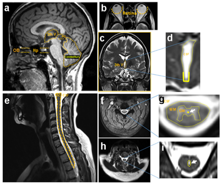
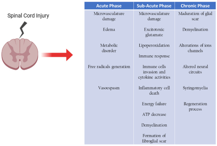
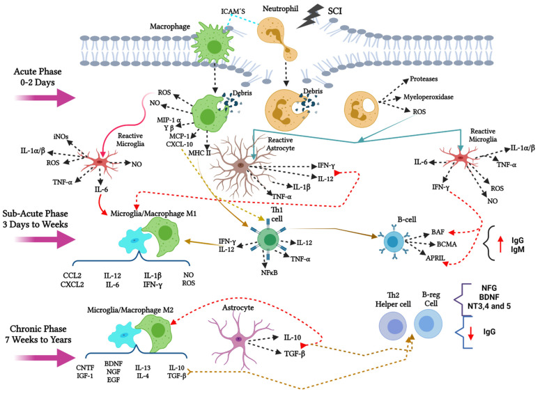
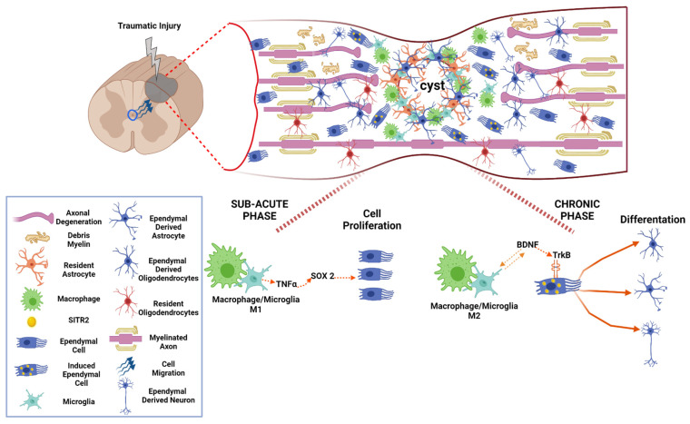
Aldynoglia are growth-promoting cells with a morphology similar to radial glia and share properties and markers with astrocytes and Schwann cells. They are distributed in several locations throughout the adult central nervous system, where the cells of the aldynoglia interact and respond to the signals of the immune cells. After spinal cord injury (SCI), the functions of resident aldynoglia, identified as ependymocytes, tanycytes, and ependymal stem cells (EpSCs) of the spinal cord are crucial for the regeneration of spinal neural tissue. These glial cells facilitate axonal regrowth and remyelination of injured axons. Here, we review the influence of M1 or M2 macrophage/microglia subpopulations on the fate of EpSCs during neuroinflammation and immune responses in the acute, subacute, and chronic phases after SCI.
Keywords: aldynoglia; axonal growth; bdnf; ensheathing cells; epscs; gfap; macrophage; microglia; p75 ngfr; sci; vimentin.
Conflict of interest statement
The authors declare no conflict of interest.
Figures
Similar articles
-
Axonal Growth and Fasciculation of Spinal Neurons Promoted by Aldynoglia in Alkaline Fibrin Hydrogel: Influence of Tol-51 Sulfoglycolipid.Int J Mol Sci. 2024 Aug 23;25(17):9173. doi: 10.3390/ijms25179173. Int J Mol Sci. 2024. PMID: 39273121 Free PMC article.
-
Glial scar and axonal regeneration in the CNS: lessons from GFAP and vimentin transgenic mice.Acta Neurochir Suppl. 2004;89:87-92. doi: 10.1007/978-3-7091-0603-7_12. Acta Neurochir Suppl. 2004. PMID: 15335106
-
Olfactory ensheathing cells: bridging the gap in spinal cord injury.Neurosurgery. 2000 Nov;47(5):1057-69. doi: 10.1097/00006123-200011000-00006. Neurosurgery. 2000. PMID: 11063098 Review.
-
Glial Cells Shape Pathology and Repair After Spinal Cord Injury.Neurotherapeutics. 2018 Jul;15(3):554-577. doi: 10.1007/s13311-018-0630-7. Neurotherapeutics. 2018. PMID: 29728852 Free PMC article. Review.
-
Glial-Neuronal Interactions in Pathogenesis and Treatment of Spinal Cord Injury.Int J Mol Sci. 2021 Dec 17;22(24):13577. doi: 10.3390/ijms222413577. Int J Mol Sci. 2021. PMID: 34948371 Free PMC article. Review.
Cited by
-
Screening the immune-related circRNAs and genes in mice of spinal cord injury by RNA sequencing.Front Immunol. 2022 Nov 21;13:1060290. doi: 10.3389/fimmu.2022.1060290. eCollection 2022. Front Immunol. 2022. PMID: 36479123 Free PMC article.
-
Mechanism Underlying Acupuncture Therapy in Spinal Cord Injury: A Narrative Overview of Preclinical Studies.Front Pharmacol. 2022 Apr 7;13:875103. doi: 10.3389/fphar.2022.875103. eCollection 2022. Front Pharmacol. 2022. PMID: 35462893 Free PMC article. Review.
-
Transplantation of Predegenerated Peripheral Nerves after Complete Spinal Cord Transection in Rats: Effect of Neural Precursor Cells and Pharmacological Treatment with the Sulfoglycolipid Tol-51.Cells. 2024 Aug 8;13(16):1324. doi: 10.3390/cells13161324. Cells. 2024. PMID: 39195214 Free PMC article.
-
From single to combinatorial therapies in spinal cord injuries for structural and functional restoration.Neural Regen Res. 2025 Mar 1;20(3):660-670. doi: 10.4103/NRR.NRR-D-23-01928. Epub 2024 Apr 16. Neural Regen Res. 2025. PMID: 38886932 Free PMC article.
-
The Promising Role of a Zebrafish Model Employed in Neural Regeneration Following a Spinal Cord Injury.Int J Mol Sci. 2023 Sep 11;24(18):13938. doi: 10.3390/ijms241813938. Int J Mol Sci. 2023. PMID: 37762240 Free PMC article. Review.
References
Publication types
MeSH terms
Grants and funding
LinkOut - more resources
Full Text Sources
Medical
Research Materials
Miscellaneous
