. 2021 Oct 22;21(1):1134.
doi: 10.1186/s12885-021-08859-5.

Antiangiogenic antibody BD0801 combined with immune checkpoint inhibitors achieves synergistic antitumor activity and affects the tumor microenvironment

Affiliations

Antiangiogenic antibody BD0801 combined with immune checkpoint inhibitors achieves synergistic antitumor activity and affects the tumor microenvironment

Liting Xue et al. BMC Cancer. .

Abstract

Background: Signaling through VEGF/VEGFR induces cancer angiogenesis and affects immune cells. An increasing number of studies have recently focused on combining anti-VEGF/VEGFR agents and immune checkpoint inhibitors (ICIs) to treat cancer in preclinical and clinical settings. BD0801 is a humanized rabbit anti-VEGF monoclonal antibody in the clinical development stage.

Methods: In this study, the anti-cancer activities of BD0801 and its potential synergistic anti-tumor effects when combined with different immunotherapies were assessed by using in vitro assays and in vivo tumor models. Ex vivo studies were conducted to reveal the possible mechanisms of actions (MOA) underlying the tumor microenvironment modification.

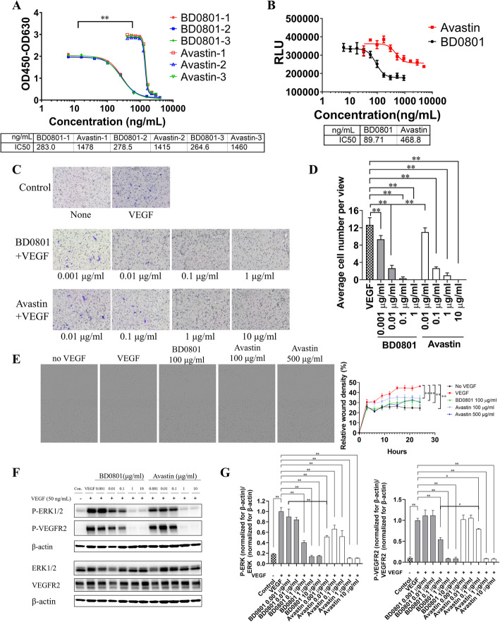
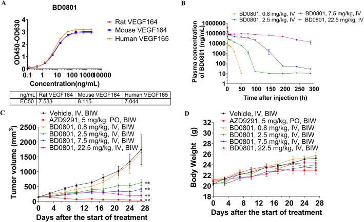
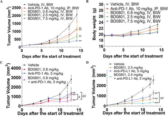
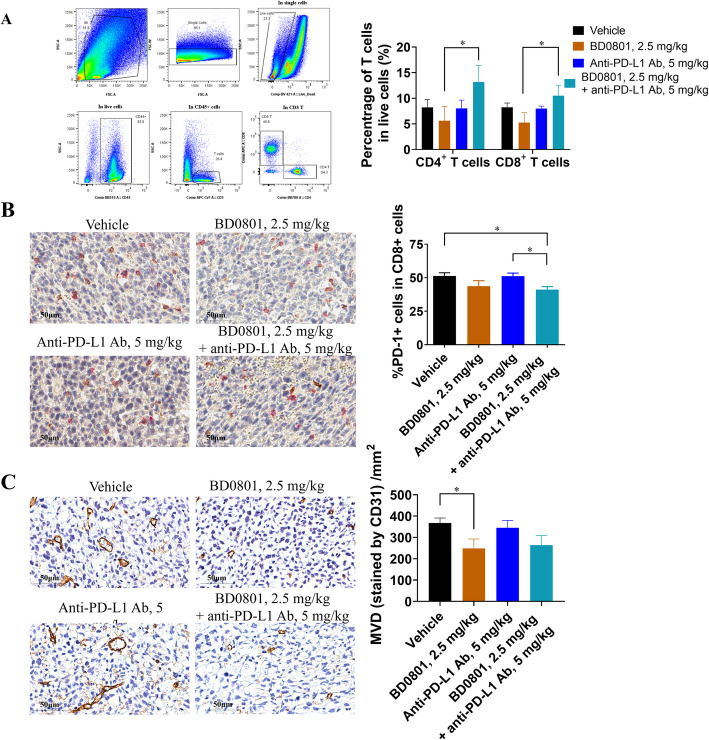
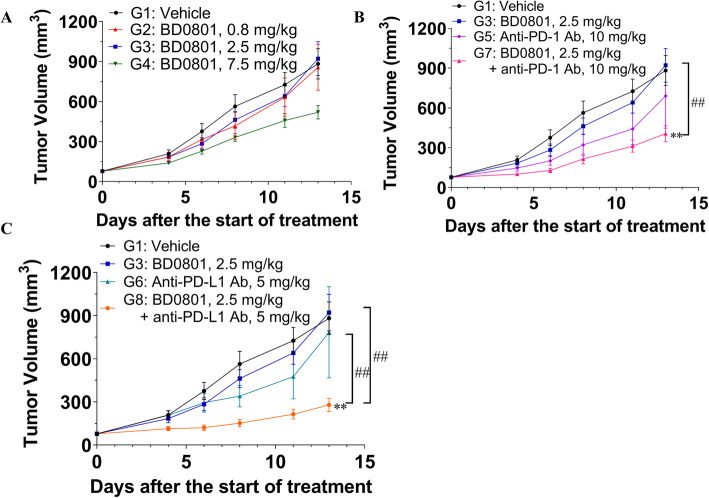
Results: BD0801 showed more potent antitumor activity than bevacizumab, reflected by stronger blockade of VEGF/VEGFR binding and enhanced inhibitory effects on human umbilical vein endothelial cells (HUVECs). BD0801 exhibited dose-dependent tumor growth inhibitory activities in xenograft and murine syngeneic tumor models. Notably, combining BD0801 with either anti-PD-1 or anti-PD-L1 antibodies showed synergistic antitumor efficacy in both lung and colorectal cancer mouse models. Furthermore, the mechanistic studies suggested that the MOA of the antitumor synergy involves improved tumor vasculature normalization and enhanced T-cell mediated immunity, including increased tumor infiltration of CD8+ and CD4+ T cells and reduced double-positive CD8+PD-1+ T cells.

Conclusions: These data provide a solid rationale for combining antiangiogenic agents with immunotherapy for cancer treatment and support further clinical development of BD0801 in combination with ICIs.

Keywords: Anti-VEGF monoclonal antibody; Antitumor synergy; Combination treatment; Immune checkpoint blockade; Tumor microenvironment.


Conflict of interest statement

The authors declare no competing interests.

Figures

Fig. 1
Fig. 1
The effects of BD0801 on VEGF/VEGFR2 binding, downstream signaling, and HUVEC cellular functions in vitro. (A) Blockade of VEGF-VEGFR2 binding by BD0801 or bevacizumab (Avastin) was analyzed by ELISA. Results of three independent experiments. (B) Different concentrations of BD0801 or bevacizumab were incubated with 50 ng/ml VEGF for 2 h at 37 °C before they were added into the HUVEC culture. After 72 h, the inhibition of HUVEC proliferation was detected by CellTiter Glo staining. The experiment was performed three times, and one of the representative experiment results is shown. RLU, Relative light unit. (C) HUVECs were cultured in the inner chamber of the Boyden Chamber Transwell. Different concentrations of BD0801 or bevacizumab were incubated with 20 ng/ml VEGF for 30 min before adding them to the outer chamber. After incubating at 37 °C for 16 h, the migrated cells were stained purple by crystal violet and observed under the microscope. Each condition was performed in triplicates. (D) Quantification of HUVEC migration assay described in (C). (E) Different concentrations of BD0801 or bevacizumab were mixed and incubated with 20 ng/ml VEGF for 30 min before they were added to starved and scratched HUVEC culture. The relative wound densities were monitored and quantified for 24 h. The representative images at 24 h are shown. (F) Different concentrations of BD0801 or bevacizumab were mixed and incubated with 50 ng/ml VEGF for 30 min before they were added to HUVEC culture for 3 min. Protein extracts were separated by Western blot for phosphorylated-ERK1/2 (P-ERK1/2), phosphorylated-VEGFR2 (P-VEGFR2), ERK1/2, and VEGFR2. The three rows above (P-ERK1/2, P-VEGFR2, and β-actin) are from Blot 1; the three rows below (ERK1/2, VEGFR2, and β-actin) are from Blot 2. This Western blot analysis was repeated independently three times, and a representative result is shown. The original uncropped blots for this particular experiment are included in Supplementary S2. (G) The quantification and statistical analysis of the three independent Western blot experiments described in (F) are included. The value for VEGF only group was normalized to one. The quantification of P-ERK1/2, P-VEGFR2, ERK1/2, and VEGFR2 normalized for β-actin respectively are included in Supplementary Figs. S3 and S4. The error bars: standard error of the mean (SEM). *, P < 0.05, **, P < 0.01
Fig. 2
Fig. 2
PK profile and antitumor effects of BD0801 in the PC9 lung cancer mouse models. (A) BD0801 has the same affinity for the rat, mouse, and human VEGF over the same concentration range. (B) PC9 cells were implanted subcutaneously into the right flank of Balb/c nude mice. When the average tumor volume reached ~ 150 mm3, the mice received a single intravenous injection of different concentrations of BD0801. Blood levels of BD0801 were analyzed by ELISA, n = 3/group. (C) Tumor volume of PC9 tumor-bearing Balb/c nude mice was administrated with the vehicle, AZD9291, or BD0801 biweekly (n = 6/group). (D) Bodyweight of PC9 tumor-bearing Balb/c nude mice was administrated with the vehicle, AZD9291, or BD0801 biweekly (n = 6/group). The error bars represent SEM. IV, intravenous; PO, oral gavage; BIW, biweekly. P-value was calculated based on the tumor volume comparing to the vehicle group using two-way ANOVA; **, P < 0.01. The tumor images are included in Supplementary Fig. S5
Fig. 3
Fig. 3
The antitumor effects of BD0801, anti-PD-1, and anti-PD-L1 antibodies in the 3LL lung cancer syngeneic mouse model. (A, B) The 3LL tumor-bearing C57BL/6 mice received the vehicle, anti-PD-1 antibody or BD0801 biweekly; the tumor volume and the bodyweight are shown in (A) and (B), respectively (n = 6/group). (C) The 3LL tumor-bearing C57BL/6 mice received 0.8 mg/kg BD0801, 5 mg/kg anti-PD-1 antibody, or combination by intraperitoneal injection biweekly (n = 10/group). (D) The 3LL tumor-bearing C57BL/6 mice received 2.5 mg/kg BD0801, 5 mg/kg anti-PD-L1 antibody, or combination by intraperitoneal injection biweekly (n = 10/group). The error bars represent SEM. IP, intraperitoneal, Ab, antibody. The P-value was calculated based on the tumor volume comparing to the assigned group using two-way ANOVA; treatment group versus vehicle group: **, P < 0.01; combination treatment group versus single treatment group: #, P < 0.05, ##, P < 0.01. The tumor images are included in Supplementary Fig. S5
Fig. 4
Fig. 4
Ex vivo analysis of tumor samples from the 3LL syngeneic mouse model. (A) Left panel, representative FACS plots and gating strategy. Right panel, bar graphs of tumor-infiltrated T cells analyzed using FACS. Percentages of CD4+ T cells (defined as Live/DeadCD45+CD3+CD4+) and CD8+ T cells (defined as Live/DeadCD45+CD3+CD8+) in total live cells (defined as Live/Dead) (n = 6/group). The representative flow images and gating strategy of 4 different groups are shown in Supplementary Fig. S7. (B) The representative images of the IHC double staining using anti-PD-1 and CD8 antibodies are shown, Red: PD-1; Brown: CD8. (left panel, scale bar: 50 μm). The average percentages of the double-positive staining of PD-1+CD8+ T cells were analyzed (right panel, n = 10/group). (C) The representative images of the IHC staining using anti-CD31 antibody are shown (left panel, scale bar: 50 μm). The CD31 IHC data were quantified (right panel, n = 6 for each group). The error bars represent SEM; *, P < 0.05
Fig. 5
Fig. 5
The antitumor effects of BD0801, anti-PD-1 antibody, and anti-PD-L1 antibody in CT26 colorectal cancer syngeneic mouse model. (A-C) The CT26 tumor-bearing Balb/c mice received BD0801, anti-PD-1 antibody, anti-PD-L1 antibody, or different combinations by intraperitoneal injection biweekly (n = 10/group). G, Group. The error bars represent SEM. The P-value was calculated based on the tumor volume comparing to the assigned group using two-way ANOVA; treatment group versus vehicle group: **, P < 0.01; combination treatment group versus single treatment group: ##, P < 0.01. The tumor images are included in Supplementary Fig. S5

References

    1. Bouis D, Kusumanto Y, Meijer C, Mulder NH, Hospers GA. A review on pro- and anti-angiogenic factors as targets of clinical intervention. Pharmacol Res. 2006;53(2):89–103. doi: 10.1016/j.phrs.2005.10.006. - DOI - PubMed
    1. Eelen G, Treps L, Li X, Carmeliet P. Basic and therapeutic aspects of angiogenesis updated. Circ Res. 2020;127(2):310–329. doi: 10.1161/CIRCRESAHA.120.316851. - DOI - PubMed
    1. Cao Y, Arbiser J, D'Amato RJ, D'Amore PA, Ingber DE, Kerbel R, Klagsbrun M, Lim S, Moses MA, Zetter B, et al. Forty-year journey of angiogenesis translational research. Sci Transl Med. 2011;3(114):114rv113. doi: 10.1126/scitranslmed.3003149. - DOI - PMC - PubMed
    1. Teleanu RI, Chircov C, Grumezescu AM, Teleanu DM. Tumor Angiogenesis and Anti-Angiogenic Strategies for Cancer Treatment. J Clin Med. 2019;9(1):84. doi: 10.3390/jcm9010084. - DOI - PMC - PubMed
    1. Ferrara N. Vascular endothelial growth factor: basic science and clinical progress. Endocr Rev. 2004;25(4):581–611. doi: 10.1210/er.2003-0027. - DOI - PubMed

Substances