. 2021 Oct 19;37(3):109846.
doi: 10.1016/j.celrep.2021.109846.

Monitoring mitochondrial calcium and metabolism in the beating MCU-KO heart

Affiliations

Monitoring mitochondrial calcium and metabolism in the beating MCU-KO heart

Anna Kosmach et al. Cell Rep. .

Abstract

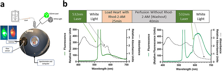
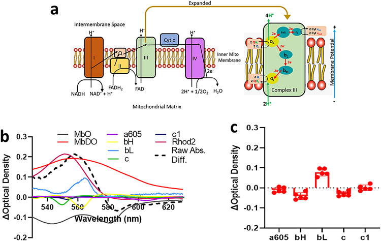
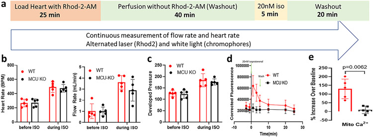
Optical methods for measuring intracellular ions including Ca2+ revolutionized our understanding of signal transduction. However, these methods are not extensively applied to intact organs due to issues including inner filter effects, motion, and available probes. Mitochondrial Ca2+ is postulated to regulate cell energetics and death pathways that are best studied in an intact organ. Here, we develop a method to optically measure mitochondrial Ca2+ and demonstrate its validity for mitochondrial Ca2+ and metabolism using hearts from wild-type mice and mice with germline knockout of the mitochondria calcium uniporter (MCU-KO). We previously reported that germline MCU-KO hearts do not show an impaired response to adrenergic stimulation. We find that these MCU-KO hearts do not take up Ca2+, consistent with no alternative Ca2+ uptake mechanisms in the absence of MCU. This approach can address the role of mitochondrial Ca2+ to the myriad of functions attributed to alterations in mitochondrial Ca2+.

Keywords: calcium; heart; isoproterenol; mitochondria; spectroscopy.


Conflict of interest statement

Declaration of interests The authors declare no competing interests.

Figures

Figure 1.
Figure 1.. Development of TOMM20-mNeonGreen tagged mouse model to confirm Rhod-2-AM loading into mitochondria.
Panel a shows fluorescence images of mNeonGreen-tagged TOMM20 in a widefield image of a WT mouse embryo (left) and a TOMM20- mNeonGreen expressing embryo (right). Panel b shows a widefield image of an embryonic mouse heart expressing mNeonGreen TOMM20. Panel c shows an excised torso section showing TOMM20-mNeonGreen expression in skeletal muscle. Scale bar represent 2 mm, 500 μm, and 2 mm for panels a-c respectively. Panel d shows Stimulated Emission Depletion (STED) microscopy image from a slice of an ex vivo mNeonGreen labelled TOMM20 (green) heart loaded with Rhod-2 (red). This image is a slice from a larger six slice z-stack that was taken over an axial range of 0.8 mm2 and deconvolved. The scale bar represents 5 μm. An enlarged image from the box region of interest is shown in panel e, and a line profile of intensities through the dotted line shown in panel f.
Figure 2.
Figure 2.. Method for interleaving fluorescent and absorbance measurements in perfused mouse heart.
Panel a shows the integrating sphere system for light collection from perfused heart. Heart is placed in chamber in center of integrating sphere, and perfusate flows outside of chamber through outflow tubing. Flow rate is collected through the flow sensor and flow box. Heart is monitored by endoscope camera. White light and 532 nm laser sources are connected to optic fiber, inserted into left ventricle of heart. Transmural light is collected through the light guide, then processed through a spectrometer and computer. Panel b shows the protocol for interleaving 532 nm laser and white light before and after Rhod-2-AM loading and wash. Left graph in panel b shows white light absorbance (dashed lines) and fluorescence emission with 532 nm laser (green) of an unloaded perfused mouse heart. Green arrows indicates 532 nm emission traces and black arrows indicate white light transmission traces. Right graph in panel b shows white light absorbance and fluorescence emission of heart following Rhod-2-AM loading and washout.
Figure 3.
Figure 3.. Correction for filtering effects.
Panel a shows fluorescence emission spectra with excitation from 532 nm laser of perfused heart before Rhod-2-AM loading (red) and the end of Rhod-2-AM washout (black). The difference spectrum between after and before Rhod-2-AM loading is shown in blue. Fluorescence spectra from Rhod-2-AM in solution taken in cuvette in integrating sphere is shown as purple dashed line. Panel b shows the raw fluorescence spectrum of a Rhod-2 loaded heart (green). Difference absorbance spectra during the hypoxia-inducing conditions of ischemia (orange) and isoproterenol (purple) compared to control are shown. The black box indicates the bandwidth of 650-680 nm in the fluorescence and difference absorbance spectra.
Figure 4.
Figure 4.. Analysis of cytochromes following Rhod-2-AM loading.
Panel a shows electron handling between cytochromes within the electron transport chain. Panel b shows the difference spectra between Rhod-2-AM washout and baseline deconvoluted using LabView program to fit reference chromophores including myoglobin (oxygenated and deoxygenated), cytochrome oxidase (a605), bH, bL, c, and c1, and Rhod-2. Panel c shows averaged peak difference absorbance for chromophores from wild-type male mice (n=5 biological replicates).
Figure 5.
Figure 5.. Mitochondrial Ca2+ measurements following isoproterenol treatment.
Panel a shows the protocol for the study. Panels b and c show heart rate (HR), flow rate (FR) and left ventricular developed pressure (LVDP) for wild-type (red) and MCU-KO (black) hearts before and during 20 nM isoproterenol treatment. Panel d shows the time course of mitochondrial Ca2+ in wild-type (red circle) and MCU-KO (black square) hearts during and following isoproterenol (ISO) treatment. N=5 biological replicates for each genotype. Panel e shows the percent changes at 1min ISO compared to baseline for mitochondrial Ca2+ in the wild-type and MCU-KO hearts.
Figure 6.
Figure 6.. Analysis of chromophores during isoproterenol treatment.
Panel a shows the time course of peak absorbance of cytochrome a605 (purple) during isoproterenol treatment and washout. Cytochrome changes were analyzed at timepoints labeled 1 (control), 2 (transient hypoxia during isoproterenol treatment), and 3 (return from hypoxia). Panels b and c show the chromophores fit using LabView showing the difference spectra between times 2 vs 1, and 3 vs 1, respectively. Panel d shows the average peak difference absorbance values from chromophores in wild-type (red) and MCU-KO (black) taken before isoproterenol treatment (time 1) and after transient hypoxia time (time 3). N=5 biological replicates for each genotype.
Figure 7.
Figure 7.. Ru360 blocks isoproterenol mediated mitochondrial Ca2+ uptake
Wild-type Langendorff hearts were treated with 2.5 ug/mL Ru360 for 10min prior to 20 nM isoproterenol treatment (n=4 biological replicates for each group). Panel a shows changes in absorbance of chromophores in heart during Ru360 treatment compared to before Ru360 (blue) compared to control hearts (red). Panel b shows heart rate during experimental protocol for control (left bars) and Ru360 treated hearts (right bars). Black bars represent heart rate for control period before Ru360 and ISO treatment. Blue bar represents heart rate during Ru360 treatment. Red bars represent heart rate during ISO treatment. Panel c shows the time course of Rhod-2 fluorescence for Ru360 treated (blue) and control (red) hearts during experimental protocol, where 20nM isoproterenol was administered at 0 min. Error bars represent SEM. Panel d shows percent increase in mitochondrial Ca2+ at 1 min ISO treatment compared to the period before ISO treatment. Panel e shows the average peak difference absorbance values from chromophores in wild-type (red) and Ru360 treated hearts (black) taken before isoproterenol treatment and after transient hypoxia.

References

    1. Aguilar-Sanchez Y, Rodriguez de Yurre A, Argenziano M, Escobar AL, and Ramos-Franco J (2019). Transmural Autonomic Regulation of Cardiac Contractility at the Intact Heart Level. Front Physiol 10, 773. - PMC - PubMed
    1. Bauer TM, Giles AV, Sun J, Femnou A, Covian R, Murphy E, and Balaban RS (2019). Perfused murine heart optical transmission spectroscopy using optical catheter and integrating sphere: Effects of ischemia/reperfusion. Anal Biochem 586, 113443. - PMC - PubMed
    1. Bisbach CM, Hutto RA, Poria D, Cleghorn WM, Abbas F, Vinberg F, Kefalov VJ, Hurley JB, and Brockerhoff SE (2020). Mitochondrial Calcium Uniporter (MCU) deficiency reveals an alternate path for Ca(2+) uptake in photoreceptor mitochondria. Sci Rep 10, 16041. - PMC - PubMed
    1. Brandes R, and Bers DM (2002). Simultaneous measurements of mitochondrial NADH and Ca(2+) during increased work in intact rat heart trabeculae. Biophys J 83, 587–604. - PMC - PubMed
    1. Chess DJ, Billings E, Covian R, Glancy B, French S, Taylor J, de BH, Murphy E, and Balaban RS (2013). Optical spectroscopy in turbid media using an integrating sphere: mitochondrial chromophore analysis during metabolic transitions. Anal Biochem. 439, 161–172. - PMC - PubMed

Publication types

MeSH terms