. 2022 Mar;87(3):1313-1328.
doi: 10.1002/mrm.29050. Epub 2021 Oct 22.

Correlated noise in brain magnetic resonance elastography

Affiliations

Correlated noise in brain magnetic resonance elastography

Ariel J Hannum et al. Magn Reson Med. 2022 Mar.

Abstract

Purpose: Magnetic resonance elastography (MRE) uses phase-contrast MRI to generate mechanical property maps of the in vivo brain through imaging of tissue deformation from induced mechanical vibration. The mechanical property estimation process in MRE can be susceptible to noise from physiological and mechanical sources encoded in the phase, which is expected to be highly correlated. This correlated noise has yet to be characterized in brain MRE, and its effects on mechanical property estimates computed using inversion algorithms are undetermined.

Methods: To characterize the effects of signal noise in MRE, we conducted 3 experiments quantifying (1) physiomechanical sources of signal noise, (2) physiological noise because of cardiac-induced movement, and (3) impact of correlated noise on mechanical property estimates. We use a correlation length metric to estimate the extent that correlated signal persists in MRE images and demonstrate the effect of correlated noise on property estimates through simulations.

Results: We found that both physiological noise and vibration noise were greater than image noise and were spatially correlated across all subjects. Added physiological and vibration noise to simulated data resulted in property maps with higher error than equivalent levels of Gaussian noise.

Conclusion: Our work provides the foundation to understand contributors to brain MRE data quality and provides recommendations for future work to correct for signal noise in MRE.

Keywords: brain; magnetic resonance elastography; physiological noise; pulsation; viscoelasticity.


Figures

Figure 1.
Figure 1.
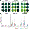
Average absolute average noise observed in one subject, demonstrating how the noise increases and spatially varies for Im, ImPhys, and ImPhysVib noise components. Similar plots for additional two subjects can be seen in Supplementary Figures S1 and S2.
Figure 2.
Figure 2.
Visual comparison of noise and noise correlation in three imaging directions. Noise is shown for one repetition of a slice alongside the corresponding correlation coefficient computed for a single point. Correlation coefficients indicate the degree of correlation of noise observations across the image with respect to the point indicated by the star.
Figure 3.
Figure 3.
Log-linear plots of correlation coefficients for a single voxel of interest indicated by the star in Figure 3. Correlation coefficients are fitted in space to determine correlation lengths. Plots demonstrate long correlation lengths in-plane versus through-plane, with reported values for each MEG direction. MEG directions are defined as x (left-right), y (anterior-posterior), and z (superior-inferior). In-plane and through-plane denominations refer to directions of correlations in space.
Figure 4.
Figure 4.
(A) Correlation lengths determined for different points in space for noise in the three MEG directions. (B) Box plots of correlation lengths from all three subjects. Top and bottom edges of box indicate 25th and 75th percentiles with center line indicating the median. Through-plane correlation lengths remain consistent across noise scans, while in-plane noise has long correlation lengths in ImPhys and ImPhysVib noise.
Figure 5.
Figure 5.
Qualitative assessments of ImPhys noise across cardiac bins and slices. (A) Average noise and (B) standard deviation of noise for one subject. There is a difference in the amount of noise between cardiac bins. Bins corresponding to systole have more noise compared to bins that correspond to the duration of diastole. This trend is consistent in all three MEG directions, with the MEG z direction having greater noise as compared to the other two directions.
Figure 6.
Figure 6.
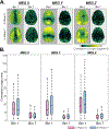
Correlation lengths differ between cardiac bin 1 and bin 7 of ImPhys noise. (A) Qualitative comparison of noise in different cardiac bins. (B) Quantitative evaluation between bin 1 and bin 7, demonstrating long correlation lengths in bin 1. Top and bottom edges of box indicate 25th and 75th percentiles and central line inside indicates median.
Figure 7.
Figure 7.
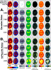
Simulation results for (A) Gaussian and (B) ImPhysVib noise. Simulated wave motion and OSS-SNR maps for datasets with noise to the simulation. Resultant estimated shear stiffness and damping ratio, and their differences to the low noise simulation. Image quality improves with gains in OSS-SNR. Results for simulations with Im and ImPhys noise added are shown in Supplementary Information (Figures S6 and S7).
Figure 8.
Figure 8.
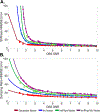
Percent error (NRMSE) in property estimates for simulations with different OSS-SNR values from Gaussian, Im, ImPhys, and ImPhysVib noise added for (A) shear stiffness and (B) damping ratio. Two-term exponential fits are included to visualize trends. Error is reduced at higher OSS-SNR levels.

References

    1. Muthupillai R, Lomas DJ, Rossman PJ, Greenleaf JF, Manduca A, Ehman RL. Magnetic resonance elastography by direct visualization of propagating acoustic strain waves. Science (80-.) 1995;269:1854–1857 doi: 10.1126/science.7569924. - DOI - PubMed
    1. Hiscox LV, Johnson CL, Barnhill E, et al. Magnetic resonance elastography (MRE) of the human brain: Technique, findings and clinical applications. Phys. Med. Biol 2016;61:R401–R437 doi: 10.1088/0031-9155/61/24/R401. - DOI - PubMed
    1. Sack I, Jöhrens K, Wuerfel J, Braun J. Structure-sensitive elastography: on the viscoelastic powerlaw behavior of in vivo human tissue in health and disease. Soft Matter 2013;9:5672–5680 doi: 10.1039/c3sm50552a. - DOI
    1. Murphy MC, Huston J, Ehman RL. MR elastography of the brain and its application in neurological diseases. Neuroimage 2019;187:176–183 doi: 10.1016/j.neuroimage.2017.10.008. - DOI - PMC - PubMed
    1. Johnson CL, Telzer EH. Magnetic resonance elastography for examining developmental changes in the mechanical properties of the brain. Dev. Cogn. Neurosci 2018;33:176–181 doi: 10.1016/j.dcn.2017.08.010. - DOI - PMC - PubMed

Publication types