Review
. 2022 Mar;41(1):53-75.
doi: 10.1007/s10555-021-09997-9. Epub 2021 Oct 23.

Glioma: molecular signature and crossroads with tumor microenvironment

Affiliations
Review

Glioma: molecular signature and crossroads with tumor microenvironment

Lennart Barthel et al. Cancer Metastasis Rev. 2022 Mar.

Abstract

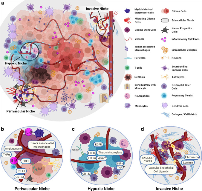
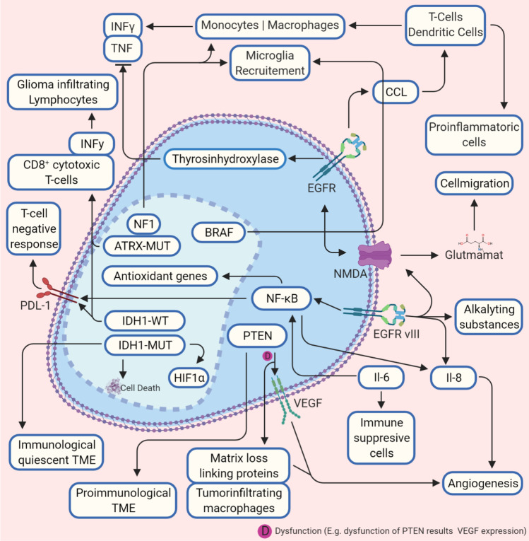
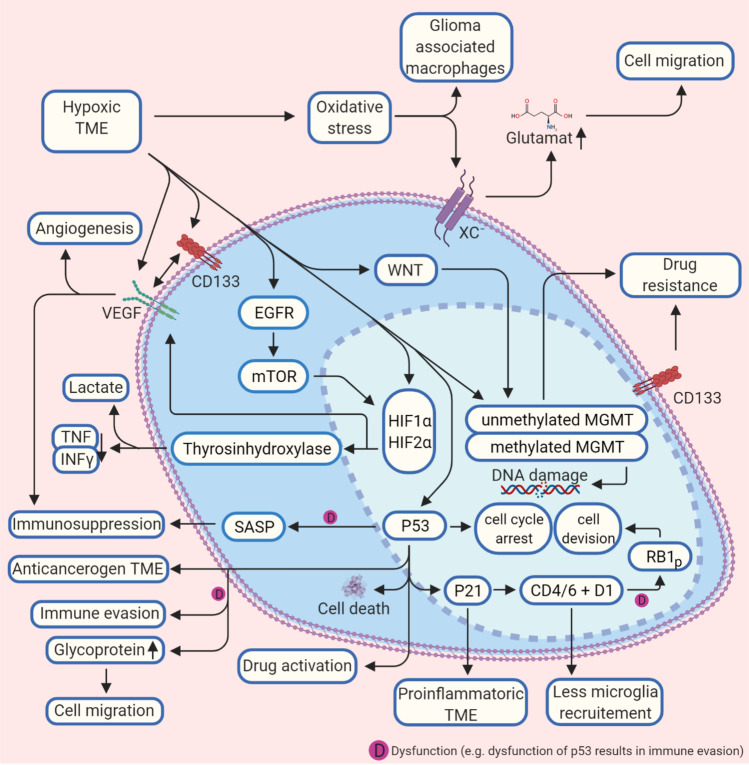
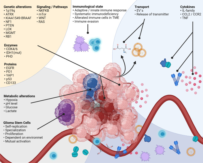
In patients with glioblastoma, the average survival time with current treatments is short, mainly due to recurrences and resistance to therapy. This insufficient treatment success is, in large parts, due to the tremendous molecular heterogeneity of gliomas, which affects the overall prognosis and response to therapies and plays a vital role in gliomas' grading. In addition, the tumor microenvironment is a major player for glioma development and resistance to therapy. Active communication between glioma cells and local or neighboring healthy cells and the immune environment promotes the cancerogenic processes and contributes to establishing glioma stem cells, which drives therapy resistance. Besides genetic alterations in the primary tumor, tumor-released factors, cytokines, proteins, extracellular vesicles, and environmental influences like hypoxia provide tumor cells the ability to evade host tumor surveillance machinery and promote disease progression. Moreover, there is increasing evidence that these players affect the molecular biological properties of gliomas and enable inter-cell communication that supports pro-cancerogenic cell properties. Identifying and characterizing these complex mechanisms are inevitably necessary to adapt therapeutic strategies and to develop novel measures. Here we provide an update about these junctions where constant traffic of biomolecules adds complexity in the management of glioblastoma.

Keywords: Cancer microenvironment; Cancer stem cells; Glioblastoma; Glioma; Stem cells; Tumor microenvironment.


Conflict of interest statement

The authors declare no competing interests.

Figures

Fig. 1
Fig. 1
The niches of GBM niches and molecular landscape. a Overview of the TME of glioblastomas, with immunological players, and the three main niches that show a certain presence of specific molecular profiles. b The vascular niche: This niche is characterized by pronounced angiogenesis with a correspondingly increased VEGF. Here tumor macrophages are accumulated. Cytokines such as IL-6 and IL-8 are increased. Likewise, PTEN leads to increased matrix cross-linking proteins, resulting in accelerated angiogenesis. c The hypoxic niche contributes to glioma growth and resistance. PTEN is increased, and HIF contributes to the upregulation of VEGF and IL-8 and supports stem cell presence indicated via increased CD133. Via tyrosine hydroxylase activity, inflammatory cytokines are reduced. d The invasive niche: This nice is marked by a normal vessel distribution and the transition to normal brain tissue. Stem cells are associated with the vessel structure, glioma cells and microglia go along in tumor growth, and glioma stem cells are associated with endothelial cells via CXCL12/CXCR4. The cellular matrix also supports invasive tumor growth (details in text). Abbreviations: CD133, CD133–prominin 1, PROM1, is a transmembrane protein; CXCL12. C-X-C motif chemokine ligand 12; CXCR4, C-X-C chemokine receptor type 4; EGFR, epidermal growth factor receptor; HIF-1α, hypoxia-inducible factor 1-alpha; HIF-2α, hypoxia-inducible factor 2-alpha; IL-6, interleukin (6); INFy, interferon gamma; MGMT, O6-methylguanine–DNA methyltransferase; PD-L1, programmed death-ligand 1; PTEN, phosphatase and tensin homolog; TNF-α, tumor necrosis factor-alpha; VEGF, vascular endothelial growth factor
Fig. 2
Fig. 2
Crossroads between molecular patterns and tumor microenvironment. A diverse mechanism influences the interactions between tumor microenvironment and heterogenous molecular parameters of glioma. In this figure, the basic interactions are shown. A pro-carcinogen tumor microenvironment is promoted by an impairment of NF-gene that leads to microglia recruitment, but also BRAF promotes recruiting via CCL2. EGFR also causes a pro-cancerogenic tumor microenvironment via CCL2 by activating monocytes, whereas the recruitment of T-cells and dendritic cells supports a pro-inflammatory tumor microenvironment. EGFRvIII also activates NMDA receptors that support cell migration through glutamate release. Via CCl2 also BRAF induces microglia recruitment. IDH1-mut and ATRX promote IFN-y and CD8+ extracellular and IDH1-mut reduce intracellular PD-L1. In contrast, IDH1-wt promotes cell death. PTEN is crucial for tumor microenvironment composition. Deficiency of PTEN leads to increased matrix cross-linking proteins, which supports angiogenesis (also via VEGF activation) and glioma migration and tumor-infiltrating macrophages. Sufficient PTEN leads to a pro-immunological tumor microenvironment (details in the text). Abbreviations: ATRX, transcriptional regulator ATRX also known as ATP-dependent helicase ATRX (-mut, mutation); BRAF, proto-oncogene B-Raf; CCL(2), CC-chemokine-ligand-(2); EGFR, epidermal growth factor receptor (vIII, variant III); IDH1, isocitrate dehydrogenase-(1) (mut, mutation; wt, wild type); HIF-1α, hypoxia-inducible factor 1-alpha; IL-6, interleukin 6; IL-8, interleukin 8; INFy, interferon gamma; NF1, neurofibromatosis type 1; NF-κB, nuclear factor “kappa-light-chain-enhancer” of activated B-cells; NMDA, N-methyl-d-aspartate; PD-L1, programmed death-ligand 1; PTEN, phosphatase and tensin homolog; TME, tumor microenvironment; TNF-(α), tumor necrosis factor (alpha); VEGF, vascular endothelial growth factor
Fig. 3
Fig. 3
Hypoxic glioblastoma tumor microenvironment and molecular interactions. A hypoxic tumor microenvironment TME influences molecular biological processes in the glioma cell on several levels. Methylation of the MGMT, VEGF, and p53-protein are activated, which reduces the effect of alkylating agents and promotes angiogenesis. p53 is also the central regulator of p21 and CDK4/D1, which reduce microglia recruitment. A malfunction of p53 leads to increased glycoprotein concentration in the TME, which supports cell migration and immune evasion, and immunosuppression (details in the text). Abbreviations: BRAF, proto-oncogene B-Raf; CD133, CD133–prominin 1, PROM1, is a transmembrane protein; CDK4/6, cyclin-dependent kinase 4 and 6; D1, cyclin D1 protein; EGFR, epidermal growth factor receptor (vIII, variant III); HIF-1α, hypoxia-inducible factor 1-alpha; HIF-2α, hypoxia-inducible factor 2-alpha; MGMT, O6-methylguanine–DNA methyltransferase; INFy, interferon gamma; mTOR, mechanistic target of rapamycin; P21, cyclin-dependent kinase inhibitor 1 or CDK-interacting protein 1; P53, tumor protein P53 or tumor suppressor p53; RB1, RB transcriptional corepressor 1; SASP, senescence-associated secretory phenotype; TME, tumor microenvironment; TNF-(α), tumor necrosis factor (alpha); VEGF, vascular endothelial growth factor; WNT, Wnt signaling pathway; xc, antiporter system xc
Fig. 4
Fig. 4
The interactions between TME and glioma cells are complex, as the multiple players of widespread origin show. Intracellular factors, pathways, cytokines, genetic alterations, or environmental properties are involved, and the molecular characteristics of glioma cells are dependent on these parameters. Furthermore, vice versa, the glioma molecular patterns influence the TME composition. The detailed interactions are listed in the text. Abbreviations: 1p19q, combined loss of the short-arm chromosome 1 (i.e., 1p) and the long-arm chromosome 19 (i.e., 19q); ATRX, transcriptional regulator ATRX also known as ATP-dependent helicase ATRX (-mut, mutation); BRAF (human gene that encodes a protein called B-Raf); CCL2, CC-chemokine-ligand-2; CCR2, C–C chemokine receptor type 2; CDK4/6, cyclin-dependent kinase 4 and 6; CD133, CD133–prominin 1, PROM1, is a transmembrane protein; EGFR, epidermal growth factor receptor (vIII, variant III); EVs, extracellular vesicles; IDH1, isocitrate dehydrogenase-(1) (mut, mutation; wt, wild type); IL-family, interleukin family; KIAA1549-BRAAF, KIAA1549 (protein-coding gene); LOX, lysyl oxidase, also known as protein-lysine 6-oxidase; MGMT, O6-methylguanine–DNA methyltransferase; mTOR, mechanistic target of rapamycin; NF1, neurofibromatosis type 1; NF-κB, nuclear factor “kappa-light-chain-enhancer” of activated B-cells; P53, tumor protein P53 or tumor suppressor p53; PD-L1, programmed death-ligand 1; PHD, prolyl hydroxylase domain enzymes; PTEN, phosphatase and tensin homolog; RAS, RAS proteins control signaling pathways that are key regulators of normal cell growth and malignant transformation; RB1, RB transcriptional corepressor 1; TME, tumor microenvironment; TNF, tumor necrosis factor; WNT, Wnt signaling pathway; antiporter system xc

References

    1. Hadjipanayis CG, Van Meir EG. Tumor initiating cells in malignant gliomas: Biology and implications for therapy. Journal of Molecular Medicine. 2009;87:363–374. doi: 10.1007/s00109-009-0440-9. - DOI - PMC - PubMed
    1. Lathia JD, Mack SC, Mulkearns-hubert EE, Valentim CLL, Rich JN. Cancer stem cells in glioblastoma. Genes and Development. 2015;29:1203–1217. doi: 10.1101/gad.261982.115.tumors. - DOI - PMC - PubMed
    1. Prager BC, Bhargava S, Mahadev V, Hubert CG, Rich JN. Glioblastoma stem cells: Driving resilience through chaos. Trends in Cancer. 2020;6(3):223–235. doi: 10.1016/j.trecan.2020.01.009. - DOI - PMC - PubMed
    1. Osuka S, Van Meir EG. Overcoming therapeutic resistance in glioblastoma: The way forward. The Journal of Clinical Investigation. 2017;127(2):415. doi: 10.1172/JCI89587. - DOI - PMC - PubMed
    1. Cole AP, Hoffmeyer E, Chetty SL, Cruz-Cruz J, C.-, Hamrick, F. H., Youssef, O., Cheshier, S., & Mitra, S. S. Microglia in the brain tumor microenvironment. Advances in experimental medicine and biology. 2020;1273:197–208. doi: 10.1007/978-3-030-49270-0_11. - DOI - PubMed

Publication types