

Giant biliary mucinous cystadenoma mimicking mesenchymal hamartoma of the liver in a child: A case report
- PMID: 34688072
- PMCID: PMC8536513
- DOI: 10.1016/j.ijscr.2021.106523
Giant biliary mucinous cystadenoma mimicking mesenchymal hamartoma of the liver in a child: A case report
Abstract
Introduction: Biliary cystadenomas (BCAs) are rare benign tumors with malignant potential. They are most commonly found in middle-aged women and are quite infrequently reported in children. Even with advanced imaging, diagnosing and distinguishing BCA from other cystic liver lesions remain challenging.
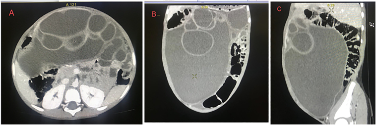
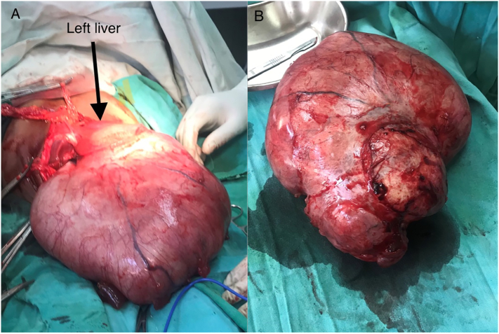
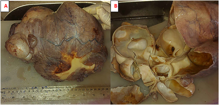
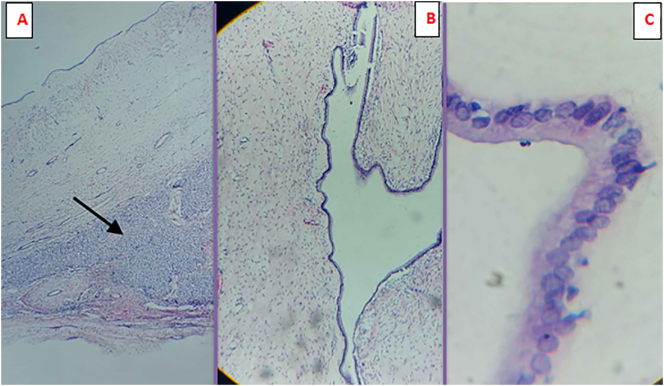
Case presentation: A 5-year-old boy was brought in by his parents to our hospital with abdominal swelling that had been persistent for a year, along with loss of appetite and weight loss. On examination, the abdomen was distended and dull on percussion. We considered mesenchymal hamartoma of the liver (MHL) as the top differential after an abdominal CT scan with contrast showed a multi-loculated cystic tumour. For both definitive diagnosis and therapy, the patient underwent exploratory laparotomy with excision of the cystic mass. Surprisingly, histopathology examination of the resected specimen revealed biliary mucinous cystadenoma (BCA).
Conclusion: Since conservative methods are associated with high recurrence rates, biliary mucinous cystic neoplasms require a high index of suspicion and should be handled with total surgical resection. In the post-operative phase, periodic surveillance imaging is recommended due to the risk of recurrence and malignant transformation.
Keywords: Biliary mucinous cystadenoma; Case report; Ethiopia; Liver cysts; Mesenchymal Hamartoma; Pediatrics.
Copyright © 2021 The Authors. Published by Elsevier Ltd.. All rights reserved.
Conflict of interest statement
None.
Figures
Similar articles
-
Biliary mucinous cystic neoplasm mimicking a hydatid cyst: a case report and literature review.BMC Gastroenterol. 2019 Jun 24;19(1):103. doi: 10.1186/s12876-019-1001-5. BMC Gastroenterol. 2019. PMID: 31234803 Free PMC article. Review.
-
Giant biliary mucinous cystadenoma of the liver.Ann Hepatol. 2013 Nov-Dec;12(6):979-83. Ann Hepatol. 2013. PMID: 24114831
-
Giant biliary cystadenoma complicated with polycystic liver: a case report.World J Gastroenterol. 2013 Oct 7;19(37):6310-4. doi: 10.3748/wjg.v19.i37.6310. World J Gastroenterol. 2013. PMID: 24115833 Free PMC article.
-
Radiologic Reporting of Simple Hepatic Cyst Versus Biliary Cystadenoma May Lead to Unnecessary Surgery.Am Surg. 2023 May;89(5):1392-1395. doi: 10.1177/00031348211054077. Epub 2021 Nov 22. Am Surg. 2023. PMID: 34806934
-
Management of Mucinous Cystic Neoplasms of the Liver.J Gastrointest Surg. 2023 Sep;27(9):1963-1970. doi: 10.1007/s11605-023-05709-6. Epub 2023 May 23. J Gastrointest Surg. 2023. PMID: 37221388 Review.
Cited by
-
Giant complex hepatic cyst causing pseudocystitis: A case report.World J Clin Cases. 2023 Nov 26;11(33):8030-8037. doi: 10.12998/wjcc.v11.i33.8030. World J Clin Cases. 2023. PMID: 38075575 Free PMC article.
-
Differentiation between mucinous cystic neoplasms and simple cysts of the liver: a systematic review and meta-analysis.Abdom Radiol (NY). 2025 Sep;50(9):4125-4138. doi: 10.1007/s00261-025-04874-3. Epub 2025 Mar 17. Abdom Radiol (NY). 2025. PMID: 40095015
References
-
- Agha R.A. The SCARE 2020 guideline: updating consensus surgical CAse REport (SCARE) guidelines. Int. J. Surg. 2020;84:226–230. - PubMed
LinkOut - more resources
Full Text Sources
