. 2022 Mar;149(3):912-922.
doi: 10.1016/j.jaci.2021.10.015. Epub 2021 Oct 22.

Upregulation of type 1 conventional dendritic cells implicates antigen cross-presentation in multisystem inflammatory syndrome

Affiliations

Upregulation of type 1 conventional dendritic cells implicates antigen cross-presentation in multisystem inflammatory syndrome

Janice J Huang et al. J Allergy Clin Immunol. 2022 Mar.

Erratum in

  • Corrigendum.
    [No authors listed] [No authors listed] J Allergy Clin Immunol. 2023 May;151(5):1414. doi: 10.1016/j.jaci.2023.03.003. J Allergy Clin Immunol. 2023. PMID: 37149373 Free PMC article. No abstract available.

Abstract

Background: Multisystem inflammatory syndrome in children (MIS-C) is an acute, febrile, severe acute respiratory syndrome coronavirus 2 (SARS-CoV-2)-associated syndrome, often with cardiohemodynamic dysfunction. Insight into mechanism of disease is still incomplete.

Objective: Our objective was to analyze immunologic features of MIS-C patients compared to febrile controls (FC).

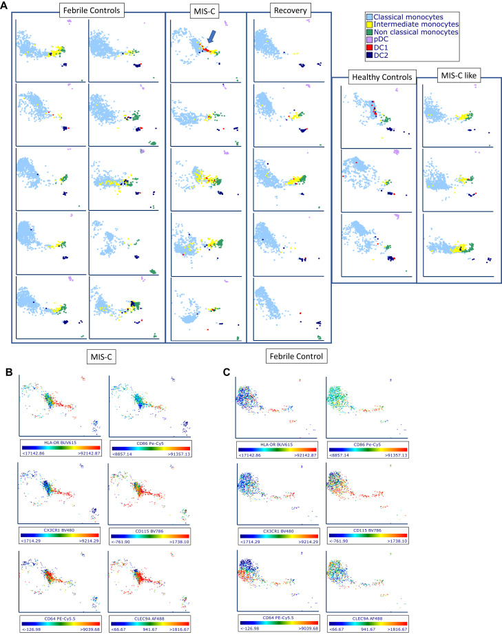
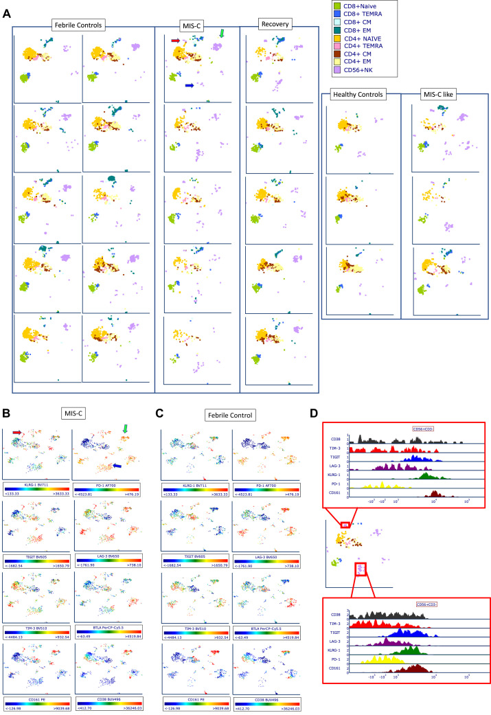
Methods: MIS-C patients were defined by narrow criteria, including having evidence of cardiohemodynamic involvement and no macrophage activation syndrome. Samples were collected from 8 completely treatment-naive patients with MIS-C (SARS-CoV-2 serology positive), 3 patients with unclassified MIS-C-like disease (serology negative), 14 FC, and 5 MIS-C recovery (RCV). Three healthy controls (HCs) were used for comparisons of normal range. Using spectral flow cytometry, we assessed 36 parameters in antigen-presenting cells (APCs) and 29 in T cells. We used biaxial analysis and uniform manifold approximation and projection (UMAP).

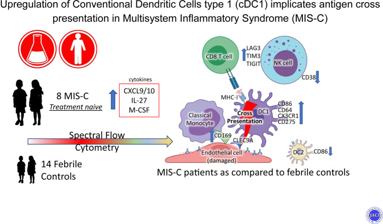
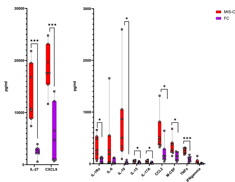
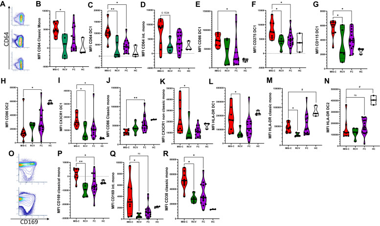
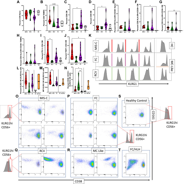
Results: Significant elevations in cytokines including CXCL9, M-CSF, and IL-27 were found in MIS-C compared to FC. Classic monocytes and type 2 dendritic cells (DCs) were downregulated (decreased CD86, HLA-DR) versus HCs; however, type 1 DCs (CD11c+CD141+CLEC9A+) were highly activated in MIS-C patients versus FC, expressing higher levels of CD86, CD275, and atypical conventional DC markers such as CD64, CD115, and CX3CR1. CD169 and CD38 were upregulated in multiple monocyte subtypes. CD56dim/CD57-/KLRGhi/CD161+/CD38- natural killer (NK) cells were a unique subset in MIS-C versus FC without macrophage activation syndrome.

Conclusion: Orchestrated by complex cytokine signaling, type 1 DC activation and NK dysregulation are key features in the pathophysiology of MIS-C. NK cell findings may suggest a relationship with macrophage activation syndrome, while type 1 DC upregulation implies a role for antigen cross-presentation.

Keywords: CLEC9A; Kawasaki disease (KD); Multisystem inflammatory syndrome in children (MIS-C); NK cell cytotoxicity; antigen cross-presentation; dendritic cells.


Figures

None
Graphical abstract
Fig 1
Fig 1
Cytokine concentrations in 7 patients with MIS-C versus 7 FC patients. IL-27 and CXCL9 are the 2 major cytokines most markedly and significantly elevated; a slight elevation but high significance is also seen in TNF-α.
Fig 2
Fig 2
Expression of markers on the APC compartment. (A) Biaxial cytometry demonstrating CD64 upregulation on CD11c+ cells in concatenated MIS-C (top), FCs (middle), and HCs (bottom). (B-D) Mean fluorescence index (MFI) of CD64 on APCs. (E-H) MFI CD86, CD275, CD115, and CX3CR1 in MIS-C versus FC and RCV. (I, J) MFI CD86 in MIS-C versus classic monocytes and DC2. (K) MFI CX3CR1 in nonclassic monocytes in MIS-C versus FC and RCV. (L-N) MFI HLA-DR expression in DC1 cells, classic monocytes, and DC2 cells. (O) Biaxial cytometry demonstrating concatenated CD169/CD38 in FC (bottom) and MIS-C (top). (P-R) MFI CD169 in classic and intermediate monocytes, and MFI CD38 in classic monocytes. ∗∗P < .01, ∗P < .05 (with multiple comparisons, Kruskal Wallis test with Dunn correction), #P < .05 (single comparisons, Mann-Whitney U test without correction); when no comparison bar is shown with HCs, a statistical test was not performed, and HC information is shown to represent an index of normal range.
Fig 3
Fig 3
UMAP analysis of APC populations. (A) Representative plots were selected from 10 FC, 5 MIS-C, 5 RCV, 5 HC, and 3 MIS-C–like subjects. MIS-C patients demonstrate small but evident population of DC1 cells concentrated at the intermediate monocyte/nonclassic monocyte location (blue arrow). This characteristic finding in our DC patients suggests that DC1 cells have altered their phenotype to take on characteristics of these monocytes, but that they are playing a unique role via the function of CLEC9A, which is typically for antigen cross-presentation. (B, C) Two selected patients, 1 with MIS-C (B) and 1 FC (C), demonstrating expression of HLA-DR, CD86, CD115, CX3CR1, CD64, and CLEC9A. Cells in the same region demonstrate upregulation of these markers, with the exception of CLEC9A, which is only seen robustly expressed in the MIS-C patient.
Fig 4
Fig 4
Expression of markers on the T and NK cell compartment. (A-D) Percentage of T-cell subtypes in patients with MIS-C versus FC and RCV. (E-H) Percentage of CD38+HLA-DR+ T-cell subsets in patients with MIS-C versus FC and RCV. (I, J) Percentage TIGIT+ and PD-1+ in CD4 T-cell CM. (K) Histogram showing upregulated KLRG1 in MIS-C patients versus RCV, HC, and FC subjects. (L) Percentage of KLRG1hi NK cells seen in MIS-C versus RCV and FC patients. (M) MFI in MIS-C patients versus RCV and FC patients; MIS-C–like patients are shown for comparison but statistical analysis not performed (line indicating where no statistical analysis performed); (N) Percentage of KLRG1hi, CD38CD161+ NK cells in MIS-C versus RCV and FC patients, MIS-C–like patients again included. (O-T) Biaxial analysis of CD38 and CD161 expression in KLRG1hi NK cells from MIS-C, FC, RCV, HC, and MIS-C–like subjects and in a FC with HLH. KLRG1hi NK cells were gated in all subgroups from the same KLRG1hi region (red box, demonstrated visually on the graph in some of the subgroups for explanation; the same region was sampled in the FC/HLH and the MIS-C–like patients). ∗∗P < .01, ∗P < .05 (with multiple comparisons, Kruskal Wallis test with Dunn correction), #P < .05 (single comparisons, Mann-Whitney U test without correction); when no comparison bar is shown with HCs, a statistical test was not performed, and HC information is shown to represent an index of normal range. HCs and MIS-C–like patients included for (M) and (N). CM, Central memory; EM, effector memory; MFI, mean fluorescence index.
Fig 5
Fig 5
UMAP analysis of T-cell and NK cell populations. (A) Ten selected FC, 5 MIS-C, 5 recovery, 5 HC, and 3 MIS-C subjects. MIS-C patients demonstrate increased number of NK cells of 2 phenotypes, indicated by a red arrow and blue arrow in the UMAP of the first MIS-C patients. These cell populations are seen also in MIS-C–like patients. MIS-C patients and FCs showed decreased expression of a third phenotype (green arrow). (B, C) Representative expression of cellular markers from 1 patient with MIS-C (B) and 1 FC (C) demonstrating KLRG1, PD-1, TIGIT, LAG3, TIM3, BTLA, CD161, and CD38. (D) Histogram of clusters indicated on previous diagrams with red arrow and blue arrow, with relative expression levels of KLRG1, TIGIT, CD161, and CD38.

Similar articles

Cited by

References

    1. Viner R.M., Whittaker E. Kawasaki-like disease: emerging complication during the COVID-19 pandemic. Lancet. 2020;395(10239):1741–1743. - PMC - PubMed
    1. Bautista-Rodriguez C., Sanchez-de-Toledo J., Clark B.C., Herberg J., Bajolle F., Randanne P.C., et al. Multisystem inflammatory syndrome in children: an international survey. Pediatrics. 2021;147 e2020024554. - PubMed
    1. Feldstein L.R., Rose E.B., Horwitz S.M., Collins J.P., Newhams M.M., Son M.B.F., et al. Multisystem Inflammatory Syndrome in U.S. Children and Adolescents. N Engl J Med. 2020;383:334–346. - PMC - PubMed
    1. Whittaker E., Bamford A., Kenny J., Kaforou M., Jones C.E., Shah P., et al. Clinical characteristics of 58 children with a pediatric inflammatory multisystem syndrome temporally associated with SARS-CoV-2. JAMA. 2020;324:259–269. - PMC - PubMed
    1. Ouldali N., Toubiana J., Antona D., Javouhey E., Madhi F., Lorrot M., et al. Association of intravenous immunoglobulins plus methylprednisolone vs immunoglobulins alone with course of fever in multisystem inflammatory syndrome in children. JAMA. 2021;325:855–864. - PMC - PubMed

Publication types

MeSH terms

Supplementary concepts