. 2021 Oct 23;7(1):310.
doi: 10.1038/s41420-021-00692-w.

Fortilin inhibits p53, halts cardiomyocyte apoptosis, and protects the heart against heart failure

Affiliations

Fortilin inhibits p53, halts cardiomyocyte apoptosis, and protects the heart against heart failure

Preedakorn Chunhacha et al. Cell Death Discov. .

Abstract

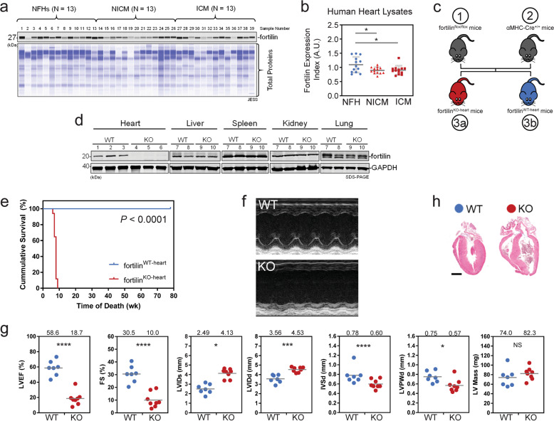
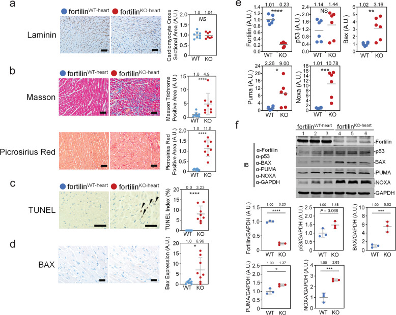
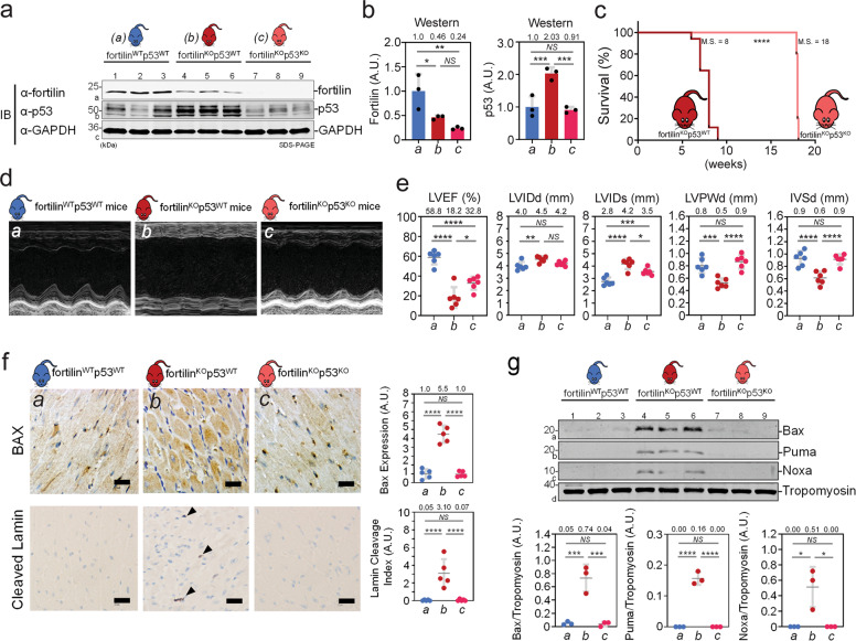
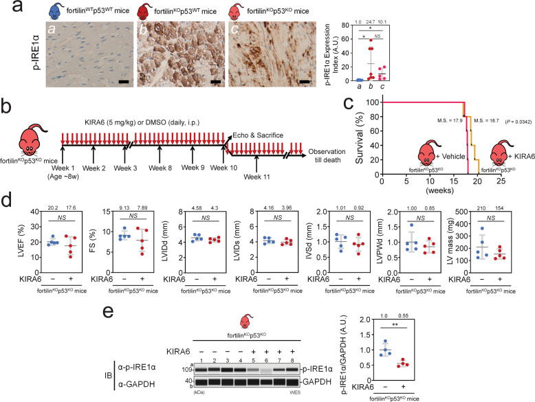
Heart failure (HF) has reached epidemic proportions in developed countries, affecting over 20 million people worldwide. Despite modern medical and device therapies, 60-70% of HF patients still die within 5 years of diagnosis as it relentlessly progresses through pervasive apoptotic loss of cardiomyocytes. Although fortilin, a 172-amino-acid anti-p53 molecule, is one of the most expressed proteins in the heart, its precise role there has remained unknown. Also unclear is how cardiomyocytes are protected against apoptosis. Here, we report that failing human hearts express less fortilin than do non-failing hearts. We also found that mice lacking fortilin in the heart (fortilinKO-heart) die by 9 weeks of age due to extensive cardiomyocyte apoptosis and severe HF, which suggests that fortilin sustains cardiomyocyte viability. The lack of fortilin is also associated with drastic upregulation of p53 target genes in the hearts. The heart-specific deletion of p53 in fortilinKO-heart mice extends their life spans from 9 to 18 weeks by mitigating cardiomyocyte apoptosis. Our data suggest that fortilin is a novel cardiac p53 inhibitor and that its inadequate expression in failing hearts and subsequent overactivation of the p53 apoptosis pathway in cardiomyocytes exacerbates HF.


Conflict of interest statement

The authors declare no competing interests.

Figures

Fig. 1
Fig. 1. Mice lacking fortilin in the heart died prematurely due to severe systolic heart failure.
a, b Fortilin expression levels were lower in the hearts of patients with NICM and ICM than in those of NFH patients. c A strain of mice lacking fortilin in the cardiomyocytes (fortilinKO-heart) (3a) and its control (fortilinWT-heart) (3b) were generated by crossing fortilinflox/flox (1) and αMHC-Cre+/+ mice (2). d Fortilin protein levels were drastically lower in the hearts of fortilinKO-heart mice than in those of fortilinWT-heart mice. e All fortilinKO-heart mice died by 9 weeks of age. f, g Transthoracic echocardiography at 7 weeks of age revealed dilated, thinned, and severely dysfunctional left ventricle in fortilinKO-heart mice. h Sagittal heart sections of fortilinWT-heart and fortilinKO-heart mice. NFH non-failing hearts, NICM non-ischemic cardiomyopathy, ICM ischemic cardiomyopathy, JESS an automated capillary-based quantitative Western blot system with in-capillary total protein evaluation capability, A.U. arbitrary units, WT fortilinWT-heart (or αMHC-Cre−/−fortilinflox/flox) mice that express fortilin normally in the heart, KO fortilinKO-heart (or αMHC-Cre+/+fortilinflox/flox) mice that do not express fortilin in the heart, GAPDH glyceraldehyde 3-phosphate dehydrogenase, LVEF left ventricular (LV) ejection fraction, FS fractional shortening, LVIDs LV internal diameters in systole, LVIDd LV internal diameters in diastole, IVSd interventricular septum thickness in diastole, LVPWd LV posterior wall thickness in diastole. Scale bar = 500 µm; Error bars, means ± SD, statistical analyses performed using Student’s two-sample t-test, NS not statistically significant; *P < 0.05; ***P < 0.005; ****P < 0.001 (see also Fig. S1).
Fig. 2
Fig. 2. Severe fibrosis and apoptosis were evident in the hearts of fortilinKO-heart mice, with signs of activation of the p53 pathway.
a The size of cardiomyocytes was similar in fortilinWT-heart and fortilinKO-heart mice, as shown in laminin staining. b Both Masson and picrosirius red staining showed severe fibrosis in the heart of fortilinKO-heart mice. c TUNEL staining revealed a significantly higher frequency of apoptotic cardiomyocytes in fortilinKO-heart hearts compared to fortilinWT-heart hearts. d BAX immunogenicity was drastically greater in fortilinKO-heart than in fortilinWT-heart mice. e The message levels of the p53-target, pro-apoptotic genes BAX, PUMA, and NOXA were significantly greater in the hearts of fortilinKO-heart mice than in those of fortilinWT-heart mice according to RT-qPCR assays of the heart RNAs. f The protein levels of BAX, PUMA, and NOXA were significantly greater in the hearts of fortilinKO-heart mice than in those of fortilinWT-heart mice, as shown by Western blot analyses of the heart lysates. A.U. arbitrary units, WT fortilinWT-heart (or αMHC-Cre−/−fortilinflox/flox) mice that express fortilin normally in the heart, KO fortilinKO-heart (or αMHC-Cre+/+fortilinflox/flox) mice, TUNEL terminal deoxynucleotidyl transferase dUTP nick-end labeling to identify apoptotic cells; scale bar = 100 µm; IB immunoblot, α-fortilin anti-fortilin antibody, GAPDH glyceraldehyde 3-phosphate dehydrogenase. Error bars means ± SD, statistical analyses performed using Student’s two-sample t-test: NS not statistically significant; *P< 0.05; **P< 0.01; ***P < 0.005, ****P < 0.001.
Fig. 3
Fig. 3. Lack of p53 partially rescued the premature death of fortilinKO-heart mice.
a, b Western blot analyses show that fortilin protein levels were significantly lower in fortilinKOp53WT and fortilinKOp53KO mice than in fortilinWTp53WT mice. p53 protein levels were significantly lower in fortilinKOp53KO mice than in fortilinKOp53WT mice. c The lack of p53 in the heart of fortilinKO-heart mice increased the M.S. from 8 to 18 weeks. d, e Echocardiography revealed that the hearts of fortilinKOp53KO mice had better overall heart function than those of fortilinKOp53WT mice, as evidenced by greater LVEF, less LVIDs (less cavity dilatation), and greater IVSd and LVPWd (less wall thinning). f Immunohistochemistry showed that the lack of p53 drastically reduced the expression levels of both BAX and cleaved lamin, an apoptosis marker. g Western blot analyses showed that the expression levels of BAX, PUMA, and NOXA were significantly decreased when p53 was deleted from fortilin-deficient hearts. a fortilinWTp53WT mice (αMHC-Cre−/−fortilinflox/floxp53flox/flox mice), b fortilinKOp53WT mice (αMHC-Cre+/+fortilinflox/floxp53WT/WT mice), c fortilinKOp53KO mice (αMHC-Cre+/+fortilinflox/floxp53flox/flox mice), IB immunoblot, α-fortilin anti-fortilin antibody, GAPDH glyceraldehyde 3-phosphate dehydrogenase, A.U. arbitrary unit, M.S. median survival, LVEF left ventricular (LV) ejection fraction, LVIDs LV internal diameters in systole, LVIDd LV internal diameters in diastole, LVPWd LV posterior wall thickness in diastole, IVSd interventricular septum thickness in diastole. Scale bar = 200 µm; Error bars, means ± SD, statistical analyses performed using ANOVA with Fisher’s multiple comparison except for the survival assay, for which the Log-Rank (Mantel–Cox) test was used: NS not statistically significant; *P < 0.05; **P < 0.01; ***P< 0.005, ****P < 0.001 (see also Figs. S2 and S3).
Fig. 4
Fig. 4. Inhibition of the IRE1α pathway by KIRA6 improved the survival of fortilinKOp53KO mice.
a Activation of the endoplasmic reticulum (ER) stress pathway, as evidenced by phosphorylation of IRE1α, occurred in the hearts of fortilinKOp53WT mice. Its activation was not fully reversed by the deletion of p53 in fortilinKOp53KO mice. b Experimental scheme. Five study and five control mice were weighed, underwent echocardiography, and were sacrificed at week 10 of treatment; the rest of the mice (N = 5 each) were observed until their deaths. c KIRA6 treatment allowed fortilinKOp53KO mice to live significantly longer than did vehicle treatment. d Echocardiography showed no significant difference between fortilinKOp53KO mice treated with either KIRA6 or vehicle. e KIRA6 treatment led to less phosphorylation and activation of IRE1α compared with vehicle treatment. a fortilinWTp53WT mice (αMHC-Cre−/−fortilinflox/floxp53flox/flox mice), b fortilinKOp53WT mice (αMHC-Cre+/+fortilinflox/floxp53WT/WT mice), c fortilinKOp53KO mice (αMHC-Cre+/+fortilinflox/floxp53flox/flox mice), p-IRE1α phosphorylated inositol-requiring enzyme 1 alpha, A.U. arbitrary unit, KIRA6 IRE1α Kinase Inhibiting RNase Attenuator 6, DMSO dimethyl sulfoxide used as vehicle, i.p. intraperitoneal injection, M.S. median survival, LVEF left ventricular (LV) ejection fraction, FS fractional shortening, LVIDd LV internal diameters in diastole, LVIDs LV internal diameters in systole, IVSd interventricular septum thickness in diastole; LVPWd LV posterior wall thickness in diastole, IB immunoblot, α-p-IRE1α anti-phosphorylated IRE1α antibody, GAPDH glyceraldehyde 3-phosphate dehydrogenase, WES an automated capillary-based quantitative Western blot system without total protein evaluation capability; Scale bar = 200 µm; Error bars, means ± SD, statistical analyses performed using Student’s two-sample t-test, except for the survival assay, for which the Log-Rank (Mantel–Cox) test was used: NS not statistically significant, *P < 0.05; **P < 0.01 (see also Fig. S4).

Similar articles

Cited by

References

    1. Mann DL. Getting pumped about heart failure. Cell Metab. 2014;19:896–7. doi: 10.1016/j.cmet.2014.05.008. - DOI - PMC - PubMed
    1. Narula J, Haider N, Arbustini E, Chandrashekhar Y. Mechanisms of disease: apoptosis in heart failure-seeing hope in death. Nat Clin Pract Cardiovasc Med. 2006;3:681–8. doi: 10.1038/ncpcardio0710. - DOI - PubMed
    1. Hartupee J, Mann DL. Neurohormonal activation in heart failure with reduced ejection fraction. Nat Rev Cardiol. 2017;14:30–8. doi: 10.1038/nrcardio.2016.163. - DOI - PMC - PubMed
    1. Guerra S, Leri A, Wang X, Finato N, Di Loreto C, Beltrami CA, et al. Myocyte death in the failing human heart is gender dependent. Circ Res. 1999;85:856–66. doi: 10.1161/01.RES.85.9.856. - DOI - PubMed
    1. Olivetti G, Abbi R, Quaini F, Kajstura J, Cheng W, Nitahara JA, et al. Apoptosis in the failing human heart. N Engl J Med. 1997;336:1131–41. doi: 10.1056/NEJM199704173361603. - DOI - PubMed