. 2022 Feb;42(2):253-263.
doi: 10.1177/0271678X211056392. Epub 2021 Oct 25.

Tenascin-C induction exacerbates post-stroke brain damage

Affiliations

Tenascin-C induction exacerbates post-stroke brain damage

Bharath Chelluboina et al. J Cereb Blood Flow Metab. 2022 Feb.

Abstract

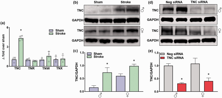
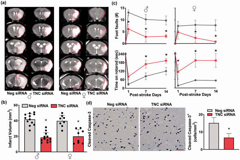
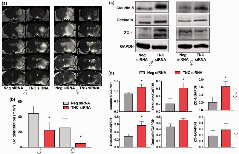
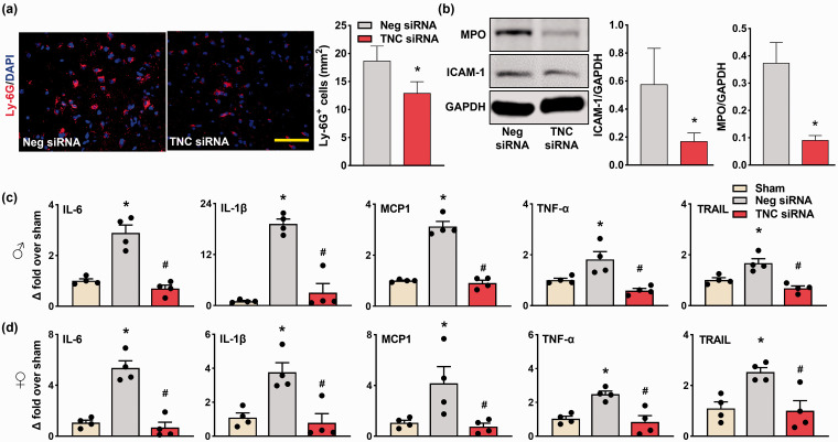
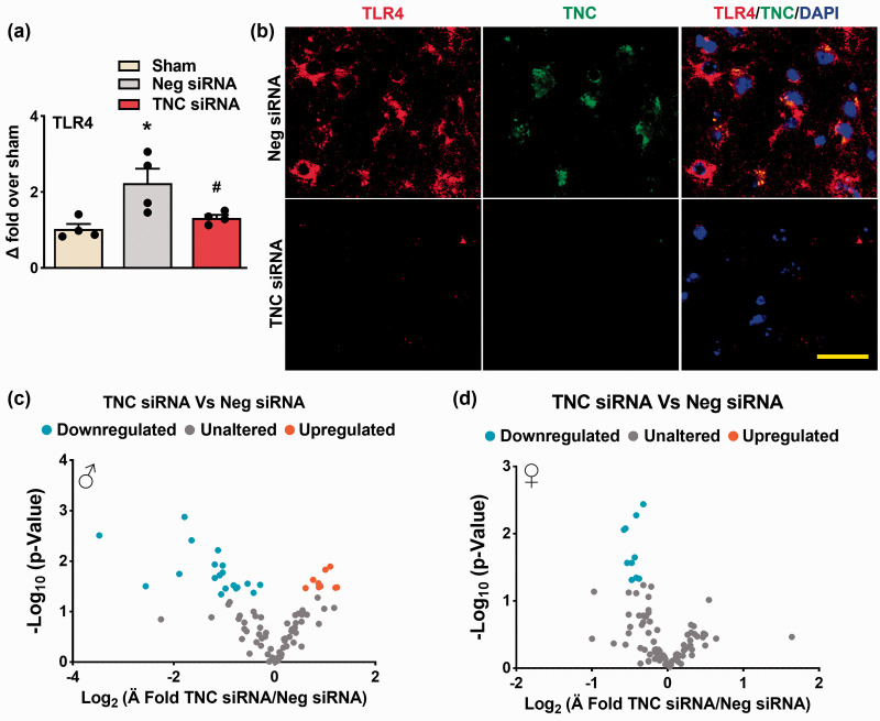
The role of tenascin-C (TNC) in ischemic stroke pathology is not known despite its prognostic association with cerebrovascular diseases. Here, we investigated the effect of TNC knockdown on post-stroke brain damage and its putative mechanism of action in adult mice of both sexes. Male and female C57BL/6 mice were subjected to transient middle cerebral artery occlusion and injected (i.v.) with either TNC siRNA or a negative (non-targeting) siRNA at 5 min after reperfusion. Motor function (beam walk and rotarod tests) was assessed between days 1 and 14 of reperfusion. Infarct volume (T2-MRI), BBB damage (T1-MRI with contrast), and inflammatory markers were measured at 3 days of reperfusion. The TNC siRNA treated cohort showed significantly curtailed post-stroke TNC protein expression, motor dysfunction, infarction, BBB damage, and inflammation compared to the sex-matched negative siRNA treated cohort. These results demonstrate that the induction of TNC during the acute period after stroke might be a mediator of post-ischemic inflammation and secondary brain damage independent of sex.

Keywords: Matricellular protein; blood-brain barrier; inflammation; ischemia-reperfusion; neuroprotection.


Conflict of interest statement

Declaration of conflicting interests: The author(s) declared no potential conflicts of interest with respect to the research, authorship, and/or publication of this article.

Figures

Figure 1.
Figure 1.
Focal ischemia significantly increased TNC protein expression, which was inhibited by TNC siRNA treatment. (a) Real-time PCR analysis shows the expression of tenascins at 24 h of reperfusion in the ipsilateral cerebral cortex of adult male mice subjected to 1 h MCAO. Values are mean ± SD; n = 4/group, *p < 0.05 compared with sham by using the Mann-Whitney U test. (b) & (c) TNC protein expression in adult male and female mice at 1 day of reperfusion following transient ischemia. (d) & (e) The TNC siRNA treated cohort showed significant knockdown compared with the non-targeting Neg siRNA treated cohort at 3 days of reperfusion following transient ischemia. The values in the histograms are means ± SD (n = 5/group). *p < 0.05 compared with the respective sham in (b) & (c) and *p < 0.05 compared with the respective Neg siRNA in (d) & (e) by using the Mann-Whitney U test.
Figure 2.
Figure 2.
TNC protein was expressed in neurons, astrocytes, and microglia after focal ischemia. Neurons, astrocytes, and microglia were immunostained with NeuN, GFAP, and TMEM119, respectively. Brain sections were from representative male and female mice of each cohort treated with TNC siRNA or Neg siRNA following 1 h of transient MCAO and 3 days of reperfusion. The scale bar is 50 μm. Similar results were observed in n = 4/group.
Figure 3.
Figure 3.
Post-stroke TNC siRNA treatment decreased brain damage and improved motor function in adult male and female mice. (a) & (b) The TNC siRNA cohort showed significantly smaller infarcts than the Neg siRNA cohort in both male and female mice subjected to 3 days of reperfusion after transient MCAO. T2 MRI scans (a) are from representative mice of the 2 groups. Bar graphs (b) values are mean ± SD (n = 12 and 8 per group for male and female, respectively). (c) Both male and female TNC siRNA cohorts (red color) showed significantly improved post-stroke motor function between days 1 and 14 of reperfusion compared with sex-matched Neg siRNA cohorts (grey color) studied by beam walk test and rotarod test. Values in the line graphs are n = 7/group for both sexes. (d) The male TNC siRNA cohort also showed a reduced number of cleaved caspase-3+ cells at 3 days of reperfusion following transient MCAO. Immunostained images are from representative mice of the 2 groups. Blue arrows indicate the cleaved caspase-3+ cells. Images are from the peri-infarct areas of the ipsilateral cortex. Values in the histogram are mean ± SD; n = 4/group. Y-axis of males and females combined (b); *p < 0.05 compared with the respective Neg siRNA cohort by using the Mann-Whitney U test (b) and (d) and two-way repeated-measures ANOVA followed by Sidak’s multiple comparisons posttest (c). The scale bar is 100 μm.
Figure 4.
Figure 4.
Post-stroke TNC siRNA treatment prevented BBB disruption. (a) & (b) T1-MRI scans and bar graphs show a reduction in Gd brain uptake in the TNC siRNA treated cohorts at 3 days reperfusion after 1 h of transient MCAO compared with the Neg siRNA cohort. Values are mean ± SD (n = 4-5/group/sex); *p < 0.05 compared with the Neg siRNA cohort by the Mann-Whitney U test. (c) & (d) Western blots and bar graphs show tight junction protein (claudin-5, occludin, and zona occludens-1) expression in the TNC siRNA or Neg siRNA treated cohorts at 3 days of reperfusion after 1 h of transient ischemia. Values are mean ± SD; Y-axis of males and females combined (b); n = 4/group, *p < 0.05, compared with the Neg siRNA by using the Mann-Whitney U test.
Figure 5.
Figure 5.
TNC knockdown prevented post-stroke inflammation. (a) The male TNC siRNA cohort showed reduced infiltration of Ly-6G+ microglia/macrophages compared with the Neg siRNA cohort. Similar staining was observed in n = 4/group. Scale bar: 100 μm. (b) The male TNC siRNA cohort showed reduced protein expression of MPO and ICAM-1 compared with the Neg siRNA cohort. (c & d) RNA levels of the inflammatory molecules (IL-6, IL-1β, MCP-1, TNF-α, and TRAIL) were significantly lower in both the male and female TNC siRNA cohorts compared with the Neg siRNA cohorts at 3 days reperfusion following transient MCAO. All parameters were estimated in the peri-infarct cortex at 3 days of reperfusion after transient MCAO. Values are mean ± SD; n = 4/group, *p < 0.05 compared with the Neg siRNA by using the Mann-Whitney U test, and */#p < 0.05 compared with the sham/Neg siRNA by one-way ANOVA with Tukey’s multiple comparisons test.
Figure 6.
Figure 6.
TNC knockdown reduced TLR-dependent post-stroke neuroinflammation. (a) TLR-4 mRNA levels were lower in the male TNC siRNA cohort compared to the Neg siRNA cohort at 3 days of reperfusion following transient ischemia. Values are mean ± SD; n = 4/group, *p < 0.05 compared with the Neg siRNA group by using the Mann-Whitney U test. (b) TNC/TLR-4 coexpression was curtailed in the male TNC siRNA treated cohort compared with the Neg siRNA cohort at 3 days reperfusion following transient MCAO. Similar results were observed with n = 4/group. Scale bar: 30 μm. (c & d) TLR signaling was curtailed by TNC knockdown after stroke in both male and female mice. n = 4/group.

References

    1. Baeten KM, Akassoglou K. Extracellular matrix and matrix receptors in blood-brain barrier formation and stroke. Dev Neurobiol 2011; 71: 1018–1039. - PMC - PubMed
    1. Chiquet-Ehrismann R, Tucker RP. Tenascins and the importance of adhesion modulation. Cold Spring Harb Perspect Biol 2011; 3: a004960–a004960. - PMC - PubMed
    1. Midwood KS, Chiquet M, Tucker RP, et al.. Tenascin-C at a glance. J Cell Sci 2016; 129: 4321–4327. - PubMed
    1. Marzeda AM, Midwood KS. Internal affairs: Tenascin-C as a clinically relevant, endogenous driver of innate immunity. J Histochem Cytochem 2018; 66: 289–304. - PMC - PubMed
    1. Clancy P, Lincz LF, Maguire J, et al.. Tenascin-C is increased in atherothrombotic stroke patients and has an anti-inflammatory effect in the human carotid artery. Biofactors 2014; 40: 448–457. - PubMed

Publication types