Review
. 2021 Oct 25;16(1):94.
doi: 10.1186/s13000-021-01151-x.

PD-L1 immunostaining: what pathologists need to know

Affiliations
Review

PD-L1 immunostaining: what pathologists need to know

Mohammed Akhtar et al. Diagn Pathol. .

Erratum in

Abstract

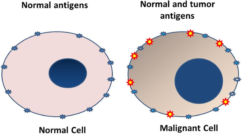
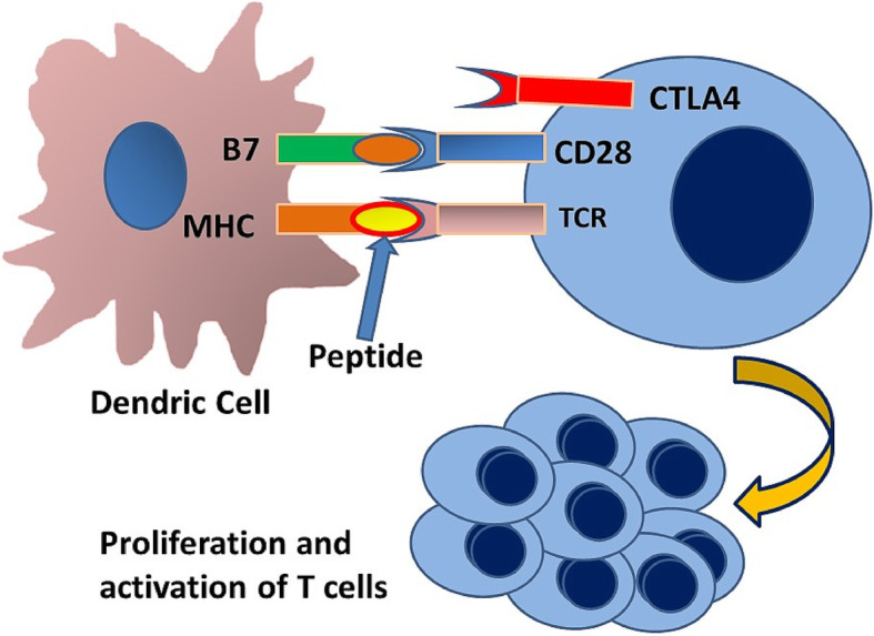
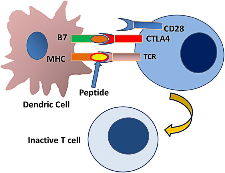
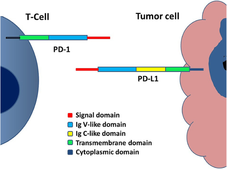
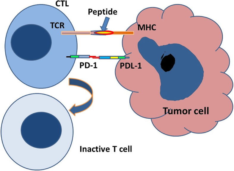
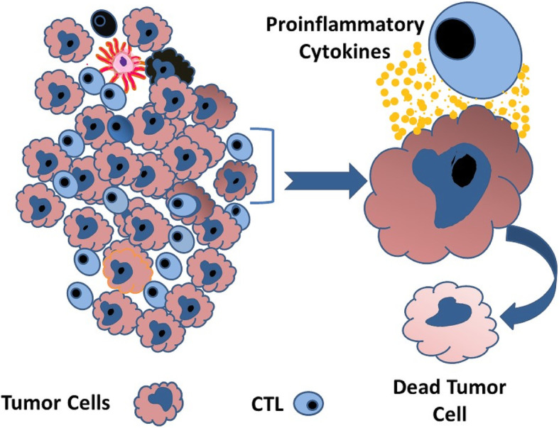
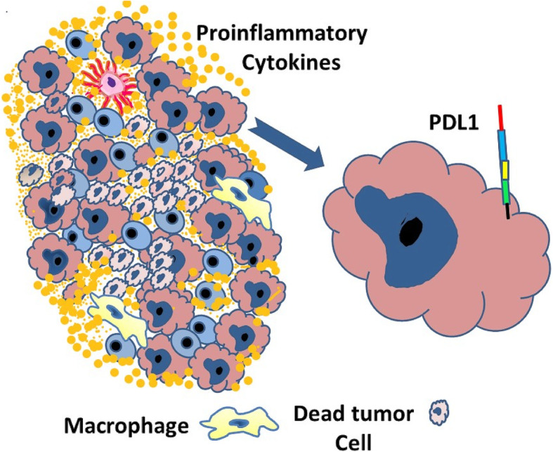
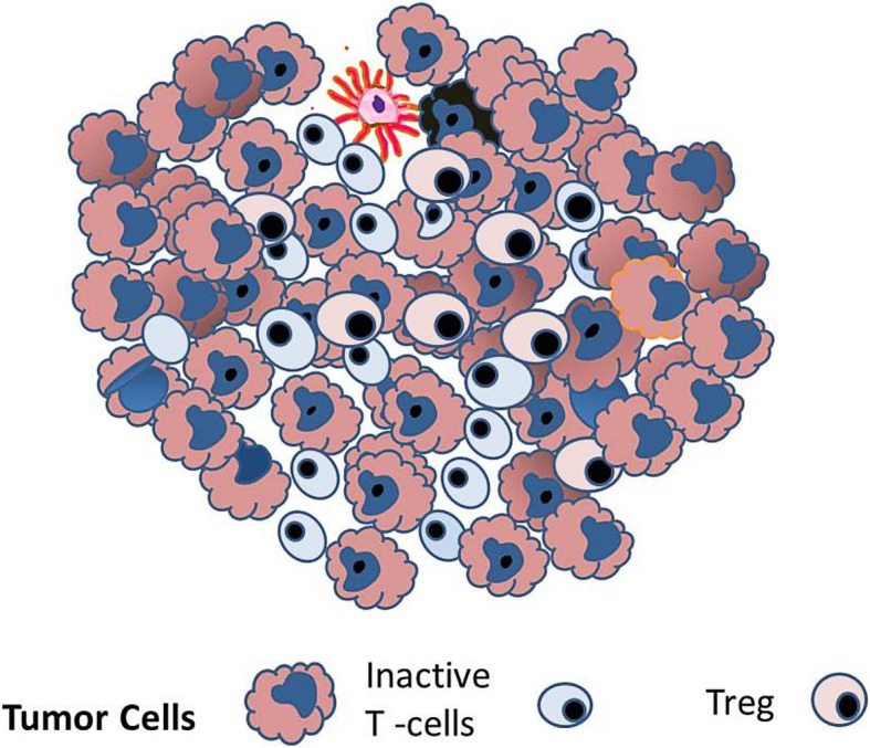
Background: Immune checkpoint proteins, especially PD-L1 and PD-1, play a crucial role in controlling the intensity and duration of the immune response, thus preventing the development of autoimmunity. These proteins play a vital role in enabling cancer cells to escape immunity, proliferate and progress.

Methods: This brief review highlights essential points related to testing for immune checkpoint therapy that histopathologists need to know.

Results: In recent years, several inhibitors of these proteins have been used to reactivate the immune system to fight cancer. Selection of patients for such therapy requires demonstration of PD-L1 activation on the tumor cells, best done by immunohistochemical staining of the tumor and immune cells using various antibodies with predetermined thresholds.

Conclusions: Immune checkpoint therapy appears to be promising and is rapidly expanding to include a large variety of cancers.

Keywords: Activation; Cancer; Immune cells; Immune checkpoint; Immunohistochemistry; Inhibition; Inhibitors; PD-1; PD-L1; T-cells.


Conflict of interest statement

The authors declare that they have no competing interests.

Figures

Fig. 1
Fig. 1
Cartoon showing surface antigens on a normal cell, compared with a cancer cell which has the normal antigens as well as additional cancer-specific and cancer-related antigen (shown as yellow-red)
Fig. 2
Fig. 2
Dendritic cells capture and process the cancer antigens and present these as peptides in association with MHC on the surface of the cells. The T-cell receptor on T- lymphocytes interacts with MHC and the peptide. In addition to binding to antigen-loaded MHC, T cells require a secondary signal to become activated. CD28 on the lymphocyte binds to the B7 on the surface of the dendritic cell. This interaction causes the T-cell to undergo stimulation and multiplication to become activated T-cells (ATC)
Fig. 3
Fig. 3
CTLA4 on the surface of ATC competes with CD28 for interaction with B7. This interaction causes the T-cell to undergo regression and inactivation
Fig. 4
Fig. 4
Cartoon revealing the structure of PD-L1 on the tumor cell and PD-1 expressed on the surface of ATC
Fig. 5
Fig. 5
Engagement of PD-L1 on the tumor cell with PD-1 on the ATC along with co-stimulation provided by T-cell receptor and MHC results in inactivation of the lymphocyte
Fig. 6
Fig. 6
Activated lymphocytes invade the tumor microenvironment and seek cancer cells for destruction. To kill the cancer cells, ATC produces pro-inflammatory cytokines, which may accumulate and alter the tumor’s micro-environment. As a result, cancer cells increase the expression of PD-L1 on the cell surface
Fig. 7
Fig. 7
Interaction of PD-L1 on cancer cells with PD-1 on the lymphocytes results in inactivation of the lymphocytes and increased numbers of Tregs ,thus providing an immune inhibitory environment
Fig. 8
Fig. 8
As a consequence of the immune inhibitory environment, cancer cells can bypass the immune system, proliferate and undergo progression. Inactivated T-cells persist and are kept in an inactive state by Tregs
Fig. 9
Fig. 9
Schematic drawing of a tumor with PD-L1 staining. There are 37 tumor cells, 14 of which are depicting membrane staining (middle part of the drawing). In addition, 10 of the tumor immune cells, including one macrophage, are positive for PD-L1 (lower right-hand corner). Based on this, tumor positive score (TPS) and combined positive score (CPS) can be calculated. formula image formula image
Fig. 10
Fig. 10
A Case of pulmonary adenocarcinoma, solid type. B Immunohistochemical staining for PD-L1 showing heterogeneous staining of tumor cells ranging from 1+ to 3+ intensity. TPS: 100. (DAKO 22C3 antibody)
Fig. 11
Fig. 11
A Case of metastatic pulmonary adenocarcinoma to the liver. B Immunohistochemical staining for tumor cells, moderate to intense staining (2+-3+) for PD-L1 TPS: 90 (DAKO 22C3 antibody)
Fig. 12
Fig. 12
A Case of buccal mucosa squamous cell carcinoma. B Immunohistochemical staining for PD-L1 revealing staining of tumor cells (right side of the figure) and tumor immune cells (left side of the figure). CPS: 90 (DAKO 22C3 antibody)
Fig. 13
Fig. 13
A Case of poorly differentiated gastric adenocarcinoma. B Immunohistochemical staining of the tumor showing staining of tumor immune cells while tumor cells are predominantly negative. CPS: 20. (DAKO 22C3 antibody)
Fig. 14
Fig. 14
A Case of moderately differentiated gastric adenocarcinoma. B immunohistochemical staining of the tumor in which only tumor cells are positive for PD-L1; while tumor immune cells are negative. CPS: 80. (DAKO 22C3 antibody)
Fig. 15
Fig. 15
A diagram depicting expression of PD-L1 positive tumor infiltrating immune cells (marked by red border) in an area of tumor cells (marked by black dotted line). The proportion of tumor area occupied by PD-L1-positive immune cells of any intensity determines the immune cell (IC) score

References

    1. Vigneron N. Human tumor antigens and cancer immunotherapy. Biomed Res Int. 2015;2015:948501. doi: 10.1155/2015/948501. - DOI - PMC - PubMed
    1. Jiang T, Shi T, Zhang H, Hu J, Song Y, Wei J, Ren S, Zhou C. Tumor neoantigens: from basic research to clinical applications. J Hematol Oncol. 2019;12(1):93. 10.1186/s13045-019-0787-5. PMID: 31492199; PMCID: PMC6731555. - PMC - PubMed
    1. Pardoll DM. The blockade of immune checkpoints in cancer immunotherapy. Nat Rev Cancer. 2012;12(4):252-64. 10.1038/nrc3239. PMID: 22437870; PMCID: PMC4856023. - PMC - PubMed
    1. He X, Xu C. Immune checkpoint signaling and cancer immunotherapy. Cell Res. 2020;30(8):660–9. doi: 10.1038/s41422-020-0343-4. - DOI - PMC - PubMed
    1. Akinleye A, Rasool Z. Immune checkpoint inhibitors of PD-L1 as cancer therapeutics. J Hematol Oncol. 2019;12(1):92. 10.1186/s13045-019-0779-5. PMID: 31488176; PMCID: PMC6729004. - PMC - PubMed

MeSH terms