Review
. 2021 Oct 8:12:723224.
doi: 10.3389/fphys.2021.723224. eCollection 2021.

Sterols, Oxysterols, and Accessible Cholesterol: Signalling for Homeostasis, in Immunity and During Development

Affiliations
Review

Sterols, Oxysterols, and Accessible Cholesterol: Signalling for Homeostasis, in Immunity and During Development

William J Griffiths et al. Front Physiol. .

Abstract

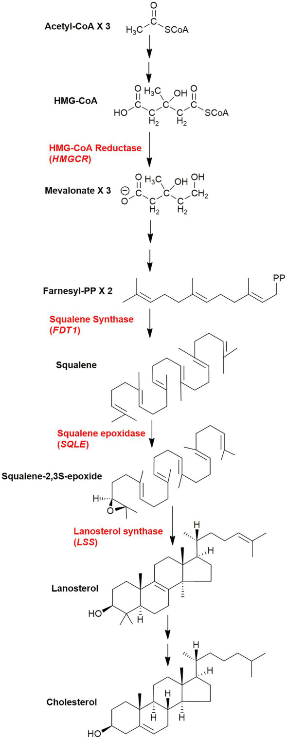
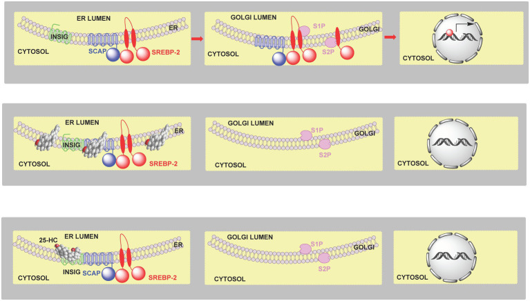
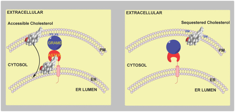
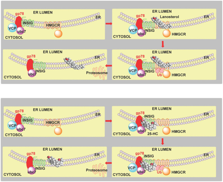
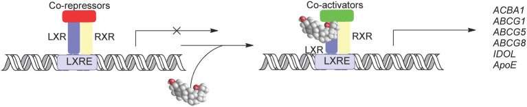
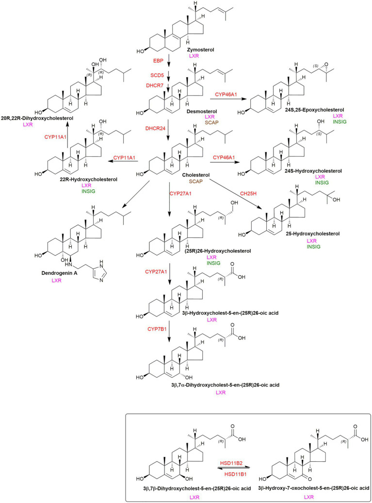
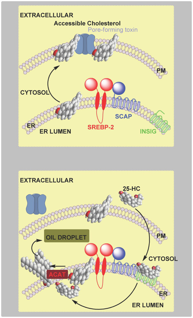
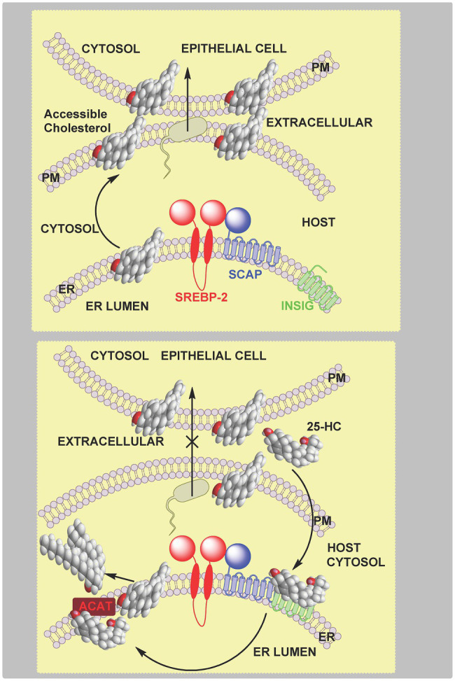
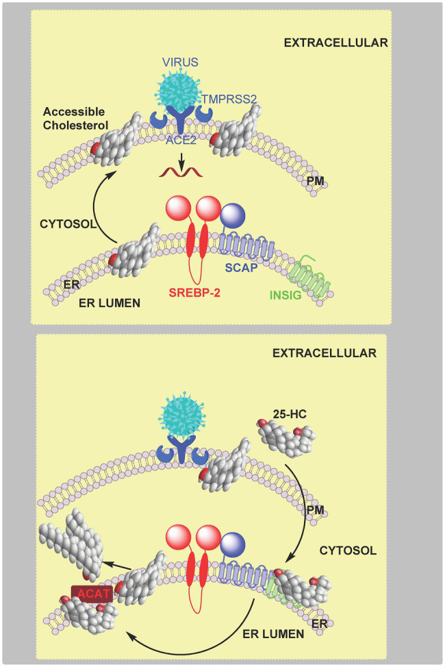
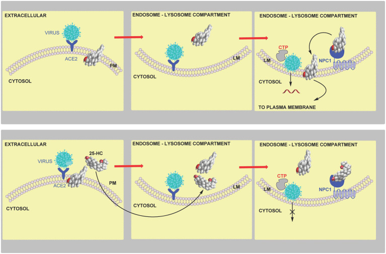
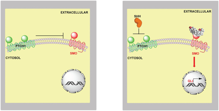
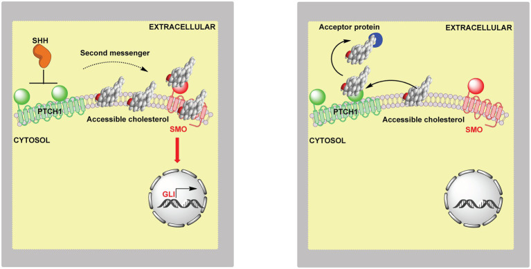
In this article we discuss the concept of accessible plasma membrane cholesterol and its involvement as a signalling molecule. Changes in plasma membrane accessible cholesterol, although only being minor in the context of total cholesterol plasma membrane cholesterol and total cell cholesterol, are a key regulator of overall cellular cholesterol homeostasis by the SREBP pathway. Accessible cholesterol also provides the second messenger between patched 1 and smoothened in the hedgehog signalling pathway important during development, and its depletion may provide a mechanism of resistance to microbial pathogens including SARS-CoV-2. We revise the hypothesis that oxysterols are a signalling form of cholesterol, in this instance as a rapidly acting and paracrine version of accessible cholesterol.

Keywords: 25-hydroxycholesterol; HMG-CoA reductase; INSIG; SARS-CoV-2; SREBP pathway; cholesterol dependent cytolysin; hedgehog signalling pathway.


Conflict of interest statement

The authors declare that the research was conducted in the absence of any commercial or financial relationships that could be construed as a potential conflict of interest.

Figures

Figure 1
Figure 1
Simplified scheme of the cholesterol biosynthetic pathway. Key enzymes are in red, genes in parenthesis. The pathway post squalene can be found in detail in Figure 6. The entire pathway including all steps and enzymes can be found in Mazein et al. (2013).
Figure 2
Figure 2
Cartoon representation of the regulation of cholesterol biosynthesis by SREBP-2. Upper panel, left to right, SCAP transports SREBP-2 from the endoplasmic reticulum to the Golgi, it is then processed to the active nuclear form which translocates to the nucleus and activates target gene transcription. Central panel, when cholesterol levels are elevated (>5 mole % endoplasmic reticulum lipids), cholesterol binds to SCAP, which then binds to INSIG retaining the SCAP-SREBP-2 complex in the endoplasmic reticulum preventing transport to, and formation of the active transcription factor, in the Golgi. Lower panel, 25-HC can bind to INSIG tethering the SCAP-SREBP-2 complex in the endoplasmic reticulum and restraining SREBP-2 transport and processing. SREBP-2 target genes include ACAT2, HMGCS, HMGCR, MVK, PMVK, MVD, IDI1, GGPS1, FDPS, FDT1, SQLE, LSS, CYP51A1, TM7SF2, MSMO1 (SC4MOL), NSDHL, HSD17B7, EBP, SC5D, DHCR7, DHCR24, LDLR, INSIG1, and STARD4 (Horton et al., 2002, 2003).
Figure 3
Figure 3
Cartoon representation of cholesterol uptake and transport from the lysosome. LDL particles bind to the LDL-receptor (LDLR) on the extracellular surface of the plasma membrane and are taken up by receptor mediated endocytosis. Endosomes combine with the lysosome where cholesterol esters are hydrolysed by LIPA. Non-esterified cholesterols is transported by soluble NPC2 and membrane bound NPC1 to the lysosomal membrane for export. Exported cholesterol is thought to travel first to the plasma membrane before reaching the endoplasmic reticulum.
Figure 4
Figure 4
Cartoon illustration of the involvement of Aster proteins in cholesterol transport and regulation. Left panel, an elevation in accessible cholesterol leads to presentation of PS on the inner leaflet of the plasma membrane and formation of a membrane contact site via the GRAM domain of the endoplasmic reticulum resident protein Aster. Accessible cholesterol is then transferred by the ASTER domain of the protein from the plasma membrane to the endoplasmic reticulum to be sensed by the SREBP-machinery. Right panel, when accessible cholesterol in the plasma membrane is below a critical level there is no binding of the Aster protein to the plasma membrane.
Figure 5
Figure 5
Lipidomoics of accessible cholesterol. Upper panel, SMase hydrolyses SM to ceramide and phosphocholine. Central panel, PTDSS1 catalyses the conversion of PC to PS. Lower panel, SPTLC2 catalyses the formation of 3-oxosphinganine from palmitoyl-CoA and serine.
Figure 6
Figure 6
Pathway from squalene to cholesterol and 24S,25-epoxycholesterol. Enzymes are indicated in red.
Figure 7
Figure 7
Cartoon representation of the sterol-induced degradation of HMG-CoA reductase. Upper panel, 4,4-dimethyl sterols induce the binding of INSIG to HMG-CoA reductase (HMGCR). The E3 ubiquitin ligase gp78 and E2 ubiquitin-conjugating enzyme ubc7 ubiquitinate (Ub) HMG-CoA reductase. In a final step stimulated by geranylgeraniol, ubiquitinated HMG-CoA reductase is extracted from the membrane by VCP and delivered to the proteosome. Lower panel, 25-HC similarly induces degradation of HMG-CoA reductase, however, it has been suggested that sterol binding is to INSIG rather than the reductase (Goldstein et al., 2006).
Figure 8
Figure 8
Activation of LXRs by oxysterol. In the absence of ligand LXR-RXR heterodimers bind to the LXR response element (LXREs) and recruit co-repressors and supress gene expression. When activated by oxysterols co-repressors are replaced by co-activators leading to the expression of LXR target genes.
Figure 9
Figure 9
Crosstalk between ligands to LXR, INSIG and SCAP. Enzymes are shown in red. LXR, INSIG and SCAP binding is illustrated where appropriate.
Figure 10
Figure 10
Cartoon representation of the protection by 25-HC of macrophages and neutrophiles against pore-forming toxins. Upper panel, pore-forming toxin binds to accessible cholesterol in the plasma membrane, oligomerises and create a pore, ultimately leading to cell death. Lower panel, 25-HC generated by macrophages in response to infection rapidly crosses the cell membrane, and (i) inhibits SREBP-2 processing leading to reduced cholesterol synthesis and (ii) activates ACAT/SOAT and cholesterol esterification, in combination leading to a reduction in plasma membrane accessible cholesterol. The consequence is reduced binding of pore-forming toxins to the plasma membrane and protection of the cell. 25-HC can also activate LXR to enhance cholesterol export and can encourage the ubiquitination and degradation of HMG-CoA reductase to repress cholesterol synthesis (not shown).
Figure 11
Figure 11
Cartoon representation of the inhibition by 25-HC of bacterial spread between epithelial cells. Upper panel, plasma membrane accessible cholesterol is required for bacterial cell–cell transmission. Lower panel, activated macrophages secrete 25-HC which stimulates the endoplasmic reticulum-located enzyme ACAT/SOAT to rapidly esterify cholesterol for storage in lipid droplets leading to reduced accessible cholesterol in the plasma membrane, this prevents bacterial spread across neighboring cells. A supporting role in reducing accessible cholesterol is played by inhibition of the SREPB-2 pathway but over a longer timeframe.
Figure 12
Figure 12
Model of coronavirus entry via the early pathway. Upper panel, virus binds to the cell surface ACE2 receptor. The membrane-bound TMPRSS2 enzyme triggers the early fusion pathway by proteolytic cleavage of the spike protein to induce the fusion-competent state of the protein. Membrane fusion proceeds and results in release of viral RNA into the cytoplasm. Membrane fusion is dependent on sufficient accessible cholesterol in the plasma membrane. Lower panel, 25-HC inhibits membrane-fusion and replication. This is achieved by reducing accessible cholesterol in the plasma membrane. Data from Wang et al. (2020) indicate that this is achieved by activating the endoplasmic reticulum enzyme ACAT/SOAT which esterifies cholesterol. An alternative explanation is 25-HC binding to INSIG and reducing cholesterol biosynthesis and up-take.
Figure 13
Figure 13
Model of coronavirus entry via the late endocytosis pathway. Upper panel, in the absence of TMPRSS2, virus is endocytosed and within the endosome-lysosome compartment low pH activates cathepsin (CTP) mediated cleavage of the spike protein triggering membrane fusion and release of viral RNA. Lower panel, 25-HC inhibits viral replication by binding to NPC1 and restricting cholesterol transport to the lysosome membrane and export, ultimately preventing membrane fusion and release of viral RNA.
Figure 14
Figure 14
Model of the involvement of oxysterols in Hh signalling. Left panel, in the absence of extracellular SHH or oxysterols PTCH1 inhibits SMO and the Hh signal is not transmitted across the membrane. Right panel, SHH relives the inhibition by PTCH1 on SMO and oxysterols bind to SMO leading to activation of GLI target genes.
Figure 15
Figure 15
Cartoon representation of the involvement of accessible cholesterol in Hh signalling. Left panel, extracellular SHH binds to PTCH1 on the cilium membrane, SMO is activated and GLI transcription factors activate GLI target genes. When SHH binds to PTCH1 it inhibits the action of PTCH1, this may lead to accumulation of accessible cholesterol which acts as the second messenger between PTCH1 and SMO. Cholesterol can bind to the CRD and TMD of SMO, binding at both sites may be required for maximal activation. Right panel, the “pump leak” model proposes PTCH1 acts as a sterol pump removing sterols from the membrane in the vicinity of SMO, thereby preventing activation of SMO by sterols. Side-chain oxysterols cross membranes far faster than cholesterol and could potentially occupy one of the sterol binding sites in activated SMO. Oxysterols can activate SMO even in the absence of the SHH ligand.

References

    1. Abdel-Khalik J., Hearn T., Dickson A. L., Crick P. J., Yutuc E., Austin-Muttitt K., et al. (2020). Bile acid biosynthesis in Smith-Lemli-Opitz syndrome bypassing cholesterol: potential importance of pathway intermediates. J. Steroid Biochem. Mol. Biol. 206:105794. doi: 10.1016/j.jsbmb.2020.105794, PMID: - DOI - PMC - PubMed
    1. Abrams M. E., Johnson K. A., Perelman S. S., Zhang L. S., Endapally S., Mar K. B., et al. (2020). Oxysterols provide innate immunity to bacterial infection by mobilizing cell surface accessible cholesterol. Nat. Microbiol. 5, 929–942. doi: 10.1038/s41564-020-0701-5, PMID: - DOI - PMC - PubMed
    1. Acton S., Rigotti A., Landschulz K. T., Xu S., Hobbs H. H., Krieger M. (1996). Identification of scavenger receptor SR-BI as a high density lipoprotein receptor. Science 271, 518–520. doi: 10.1126/science.271.5248.518, PMID: - DOI - PubMed
    1. Babiker A., Andersson O., Lindblom D., van der Linden J., Wiklund B., Lutjohann D., et al. (1999). Elimination of cholesterol as cholestenoic acid in human lung by sterol 27-hydroxylase: evidence that most of this steroid in the circulation is of pulmonary origin. J. Lipid Res. 40, 1417–1425. doi: 10.1016/S0022-2275(20)33383-6, PMID: - DOI - PubMed
    1. Bauman D. R., Bitmansour A. D., McDonald J. G., Thompson B. M., Liang G., Russell D. W. (2009). 25-Hydroxycholesterol secreted by macrophages in response to toll-like receptor activation suppresses immunoglobulin A production. Proc. Natl. Acad. Sci. U. S. A. 106, 16764–16769. doi: 10.1073/pnas.0909142106, PMID: - DOI - PMC - PubMed