. 2021 Oct 8:12:731026.
doi: 10.3389/fmicb.2021.731026. eCollection 2021.

Full-Length Galectin-3 Is Required for High Affinity Microbial Interactions and Antimicrobial Activity

Affiliations

Full-Length Galectin-3 Is Required for High Affinity Microbial Interactions and Antimicrobial Activity

Shang-Chuen Wu et al. Front Microbiol. .

Abstract

While adaptive immunity enables the recognition of a wide range of microbial antigens, immunological tolerance limits reactively toward self to reduce autoimmunity. Some bacteria decorate themselves with self-like antigens as a form of molecular mimicry to limit recognition by adaptive immunity. Recent studies suggest that galectin-4 (Gal-4) and galectin-8 (Gal-8) may provide a unique form of innate immunity against molecular mimicry by specifically targeting microbes that decorate themselves in self-like antigens. However, the binding specificity and antimicrobial activity of many human galectins remain incompletely explored. In this study, we defined the binding specificity of galectin-3 (Gal-3), the first galectin shown to engage microbial glycans. Gal-3 exhibited high binding toward mammalian blood group A, B, and αGal antigens in a glycan microarray format. In the absence of the N-terminal domain, the C-terminal domain of Gal-3 (Gal-3C) alone exhibited a similar overall binding pattern, but failed to display the same level of binding for glycans over a range of concentrations. Similar to the recognition of mammalian glycans, Gal-3 and Gal-3C also specifically engaged distinct microbial glycans isolated and printed in a microarray format, with Gal-3 exhibiting higher binding at lower concentrations toward microbial glycans than Gal-3C. Importantly, Gal-3 and Gal-3C interactions on the microbial microarray accurately predicted actual interactions toward intact microbes, with Gal-3 and Gal-3C displaying carbohydrate-dependent binding toward distinct strains of Providentia alcalifaciens and Klebsiella pneumoniae that express mammalian-like antigens, while failing to recognize similar strains that express unrelated antigens. While both Gal-3 and Gal-3C recognized specific strains of P. alcalifaciens and K. pneumoniae, only Gal-3 was able to exhibit antimicrobial activity even when evaluated at higher concentrations. These results demonstrate that while Gal-3 and Gal-3C specifically engage distinct mammalian and microbial glycans, Gal-3C alone does not possess antimicrobial activity.

Keywords: antimicrobial; blood group; galectin; microbe; molecular mimicry.


Conflict of interest statement

The authors declare that the research was conducted in the absence of any commercial or financial relationships that could be construed as a potential conflict of interest.

Figures

Figure 1
Figure 1
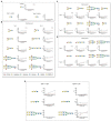
Gal-3 and Gal-3C preferentially recognize blood group antigens at distinct concentrations. Consortium for functional glycomics (CFG) glycan microarray data obtained after incubation with the indicated concentrations of Gal-3 (A) and Gal-3C (B). RFU, relative fluorescence units. Error bars represent means ± standard deviation (SD).
Figure 2
Figure 2
Gal-3 displays high affinity for blood group antigens on a CFG glycan microarray. (A-E) Binding isotherms generated following incubation of Gal-3 (blue) and Gal- 3C (red) on the CFG glycan microarray are shown for lactose and N-acetyllactosamine (LacNAc) (A), blood group A containing glycan structures (B), blood group B containing glycan structures (C), H antigen (blood group O) containing glycan structures (D) and -Gal containing glycan structures (E). Detailed Symbol Nomenclature for Glycans (SNFG) structures are shown. Error bars represent means ± standard deviation (SD). GalNAc: N-Acetylgalactosamine; GlcNAc: N-Acetylglucosamine.
Figure 3
Figure 3
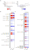
KD values for Gal-3 and Gal-3C binding toward blood group antigens and other mammalian glycans. (A) Representative binding isotherms used to generate KD values and the % max of the highest concentration tested for unsaturated glycans. (B,C) Selected blood group antigens were shown along with heat map representation of KD values (red) and the % max of the highest concentration tested for unsaturated glycans (blue) for antigens located on N-glycans (B) and non-N-glycans (C). The heat map from darker red (low KD) to light red (high KD) is shown. For the unsaturated binding, the heat map from light blue (low % max) to darker blue (high % max) is shown. Examples of glycans examined are annotated to the left of each heat map as structures that are present on N-glycans (N-glycans) as shown in (B) or as the isolated glycan motifs (non-N-glycans) as shown in (C).
Figure 4
Figure 4
Gal-3 and Gal-3C recognize distinct microbial glycans (A,B) Microbial glycan microarray (MGM) data obtained after incubation with the Gal-3 (A) or Gal-3C (B) at the concentrations indicated. Error bars represent means ± standard deviation (SD). RFU, relative fluorescence units; PA O5, Providencia alcalifaciens O5; PA O6, P. alcalifaciens O6; KP O1, Klebsiella pneumoniae O1; SP 14, Streptococcus pneumoniae type 14; SP 43, S. pneumoniae type 43; SP54, S. pneumoniae type 54; PV O45, Proteus vulgaris O45; PV O47, P. vulgaris O47; EC O55, Escherichia coli O55.
Figure 5
Figure 5
Gal-3 and Gal-3C exhibit high affinity interactions with select microbial glycans. The binding isotherms of Gal-3 (blue) and Gal-3C (red) for microbial glycans are shown. The structure for each corresponding glycan is depicted on the left of each binding isotherm. Symbols used to represent each monosaccharide present in each bacterial glycan are represented in the legend below. Error bars represent means ± standard deviation (SD). GlcA: D-Glucuronic acid, GalA: D-Galacturonic acid and ManNAc: N-Acetyl-D-mannosamine
Figure 6
Figure 6
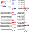
KD values of Gal-3 and Gal-3C binding to microbial glycans. Selected bacteria are presented along with heat map representation of KD and unsaturated binding (% max). The KD and % max values were sorted by red and blue color, respectively. The heat map from darker red (low KD) to light red (high KD). For the unsaturated binding, the heat map from light blue (low % max) to darker blue (high % max).
Figure 7
Figure 7
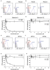
Gal-3 and Gal-3C recognize and kill Providencia alcalifaciens O5 (PA O5) and Klebsiella pneumoniae O1 (KP O1). (A,B) Flow cytometric analysis of Gal-3 (A) and Gal-3C (B) binding to PA O5 and PA O21 with or without inclusion of 20mM thiodigalactoside (TDG) as indicated. (C,D) Quantification of viable bacteria after incubation with the indicated concentrations Gal-3 (C) and Gal-3C (D). (E,F) Flow cytometric analysis of Gal-3 (E) and Gal-3C (F) binding to KP O1 and KP O4 with or without inclusion of 20mM TDG as indicated. (G,H) Quantification of viable bacteria after incubation with the indicated concentrations Gal-3 (G) and Gal-3C (H). Error bars represent means ± SD.

References

    1. Ahmad N., Gabius H. J., Andre S., Kaltner H., Sabesan S., Roy R., et al. . (2004a). Galectin-3 precipitates as a pentamer with synthetic multivalent carbohydrates and forms heterogeneous cross-linked complexes. J. Biol. Chem. 279, 10841–10847. doi: 10.1074/jbc.M312834200, PMID: - DOI - PubMed
    1. Ahmad N., Gabius H. J., Sabesan S., Oscarson S., Brewer C. F. (2004b). Thermodynamic binding studies of bivalent oligosaccharides to galectin-1, galectin-3, and the carbohydrate recognition domain of galectin-3. Glycobiology 14, 817–825. doi: 10.1093/glycob/cwh095, PMID: - DOI - PubMed
    1. Arthur C. M., Baruffi M. D., Cummings R. D., Stowell S. R. (2015a). Evolving mechanistic insights into galectin functions. Methods Mol. Biol. 1207, 1–35. doi: 10.1007/978-1-4939-1396-1_1, PMID: - DOI - PMC - PubMed
    1. Arthur C. M., Cummings R. D., Stowell S. R. (2015b). Evaluation of the bactericidal activity of galectins. Methods Mol. Biol. 1207, 421–430. doi: 10.1007/978-1-4939-1396-1_27, PMID: - DOI - PMC - PubMed
    1. Arthur C. M., Patel S. R., Mener A., Kamili N. A., Fasano R. M., Meyer E., et al. . (2015c). Innate immunity against molecular mimicry: Examining galectin-mediated antimicrobial activity. BioEssays 37, 1327–1337. doi: 10.1002/bies.201500055, PMID: - DOI - PMC - PubMed

LinkOut - more resources