. 2021 Oct 8:12:710372.
doi: 10.3389/fimmu.2021.710372. eCollection 2021.

Plasma EVs Display Antigen-Presenting Characteristics in Patients With Allergic Rhinitis and Promote Differentiation of Th2 Cells

Affiliations

Plasma EVs Display Antigen-Presenting Characteristics in Patients With Allergic Rhinitis and Promote Differentiation of Th2 Cells

Shu-Bin Fang et al. Front Immunol. .

Abstract

Background: Allergic rhinitis (AR) is characterized by IgE-mediated mucosa response after exposure to allergens. Extracellular vesicles (EVs) are nano-size vesicles containing biological cargos for intercellular communications. However, the role of plasma EVs in pathogenesis of AR remains largely unknown.

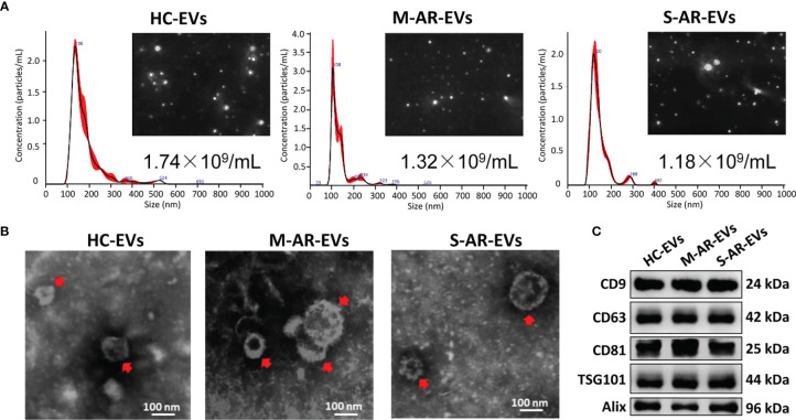
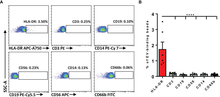
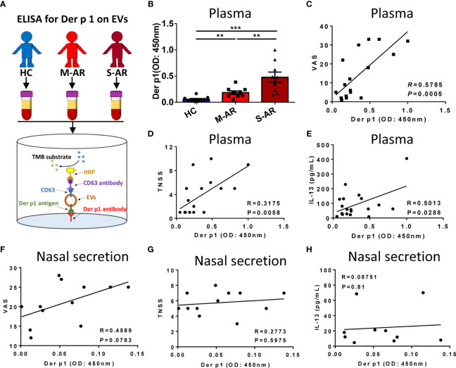
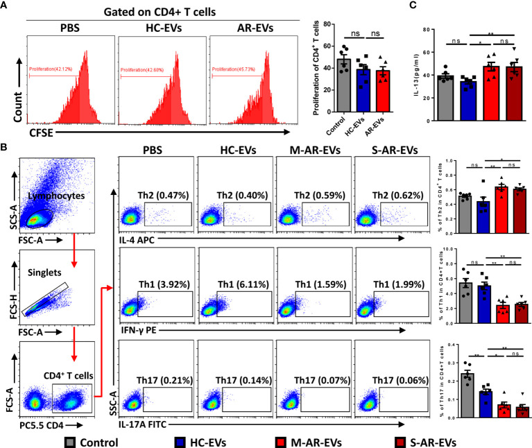
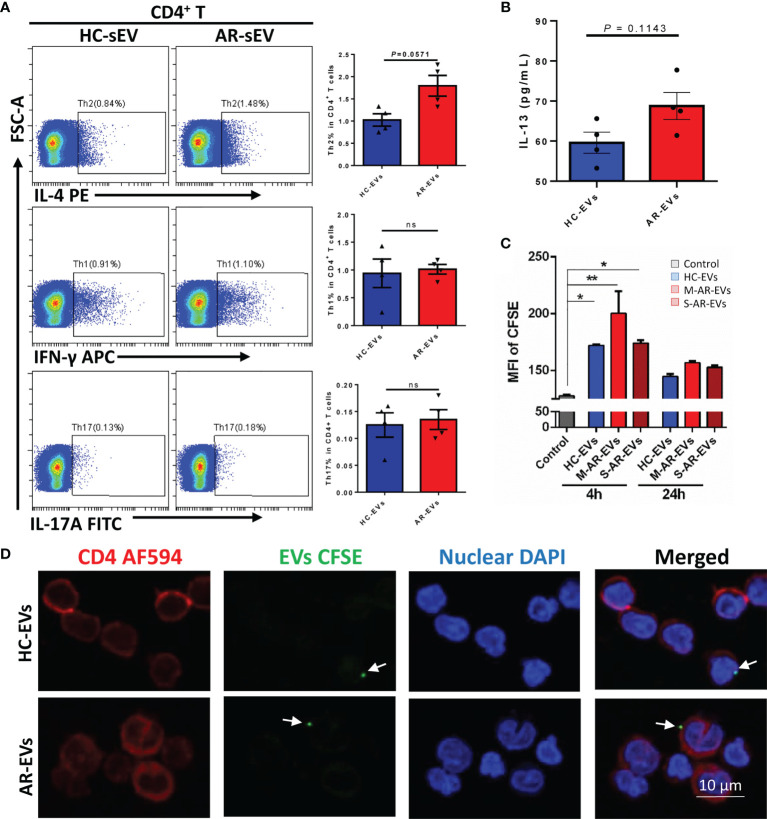
Methods: Plasma EVs from patients with AR were isolated, quantified, and characterized. The expression of Der p 1 and antigen-presenting molecules on EVs was determined by Western blot, flow cytometry, or ELISA. PKH26- and CFSE (carboxyfluorescein succinimidyl ester)-stained AR-EVs were used to determine the uptake of EVs by CD4+T cells and their effects on CD4+T cell proliferation, respectively.

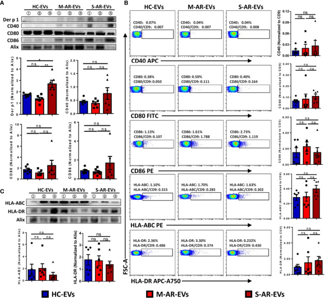
Results: Plasma EVs in healthy control (HC) and AR patients were similar in the concentration of particles, expression for specific EV markers, and both had structural lipid bilayer. However, the levels of Der p 1 on plasma EVs from both mild and moderate-severe AR patients were significantly higher than that on HC. The levels of antigen-presenting molecules on plasma EVs were similar from three subjects. Moreover, levels of Der p 1 on EVs in plasma, but not nasal secretion, were significantly associated with the symptom score of AR patients and level of plasma IL-13. Additionally, plasma EVs from patients with AR promoted the development of Th2 cells, while no effect was found on CD4+ T-cell proliferation.

Conclusions: Plasma EVs derived from patients with AR exhibited antigen-presenting characteristics and promoted differentiation of Th2 cells, thus providing novel understanding of the pathogenesis of AR.

Keywords: Der p 1; allergic rhinitis; antigen presentation; extracellular vesicles; type 2 T helper cells.


Conflict of interest statement

The authors declare that the research was conducted in the absence of any commercial or financial relationships that could be construed as a potential conflict of interest.

Figures

Figure 1
Figure 1
Characterization of plasma EVs. (A) Nanoparticle tracking analysis for determination of the particle numbers and size distribution of plasma EVs. (B) Transmission electron microscopy for characterization of the morphology of plasma EVs. (C) Plasma EVs isolated from HC subjects and patients with M-AR and S-AR were positive in the expression of specific EV markers. HC, Healthy control; M-AR, Mild allergic rhinitis; S-AR, Moderate-severe allergic rhinitis.
Figure 2
Figure 2
Determination of the immunocyte-associated markers on plasma EVs of AR patients. (A) Representative dot plots for flow cytometry analysis of immunocyte-related markers on plasma EVs. (B) Flow cytometry analysis of the expression of immunocyte-related markers on plasma EVs. Data are shown as mean ± SEM. ****p < 0.0001 by one-way ANOVA multiple comparison.
Figure 3
Figure 3
Determination of Der p 1, costimulatory molecules, and antigen presentation molecules on plasma EVs. The expression of costimulatory molecules (A) and antigen presentation molecules (B) was similar among plasma EVs derived from HC subjects and patients with M-AR and S-AR, while the expression of Der p 1 was significantly higher in patients with S-AR, as determined by Western blot. (C) No significant difference was found on levels of costimulatory and antigen presentation molecules on plasma EVs derived from HC subjects and patients with M-AR and S-AR, as suggested by flow cytometry analysis. EVs, Extracellular vesicles; HC, Healthy control; M-AR, Mild allergic rhinitis; ns, no significance; S-AR, Moderate-severe allergic rhinitis. Data are shown as mean ± SEM. Western blot of Der p 1, CD40, CD80, and CD86, as well as flow cytometry analysis of CD86 and HLA-ABC, were analyzed by Mann–Whitney U test; the other data were analyzed by one-way ANOVA multiple comparison. *p < 0.05. The data were combined from two independent experiments.
Figure 4
Figure 4
Detection of Der p 1-carrying EVs and the correlation between Der p 1+ EVs with the signs of the diseases and level of IL-13 in AR patients. (A) Schematic diagram of the ELISA for detection of Der p 1 on plasma EVs. (B) Levels of Der p 1 in patients with M-AR and S-AR were both significantly higher than HC subjects, as determined by ELISA. (C–E) Level of plasma EV-bearing Der p 1 in AR patients was significantly correlated with the VAS score (C), TNSS score (D), and level of IL-13 in plasma (E). (F–H) Level of Der p 1 on nasal secretion-derived EVs in AR patients has no significant correlation with VAS score (F), TNSS score (G), and level of IL-13 in nasal secretion (H). ELISA, Enzyme-linked immunosorbent assay; EVs, Extracellular vesicles; HC, Healthy control; M-AR, Mild allergic rhinitis; S-AR, Moderate-severe allergic rhinitis; TNSS, Total nasal symptom score; VAS, Visual analogue scale. **p < 0.01, ***p < 0.001 by one-way ANOVA multiple comparison. Correlation analyses were performed using Pearson correlation coefficient.
Figure 5
Figure 5
Effects of plasma EVs isolated from AR patients on the differentiation of T helper cells in PBMCs. (A) Flow cytometry analysis showed that no significant difference was observed on the proliferation of CD4+ T cells after treated with HC-EVs and AR-EVs. (B) Level of Th2 was increased in PBMCs treated with M-AR-EVs and S-AR-EVs compared with those treated with HC-EVs, while levels of Th1 and Th17 were oppositely reduced by M-AR-EVs and S-AR-EVs. (C) Level of IL-13 was significantly increased in the supernatants of AR-EV-stimulated PBMCs. EVs, Extracellular vesicles; HC, Healthy control; M-AR, Mild allergic rhinitis; S-AR, Moderate-severe allergic rhinitis; Th1, type 1 T helper cells; Th2, type 2 T helper cells; Th17, type 17 T helper cells; ns, no significance. Flow cytometry analyses of T helper cells and ELISA for IL-13 were analyzed by Mann–Whitney U test. Flow cytometry analyses of proliferation were analyzed by one-way ANOVA multiple comparison. *p < 0.05, **p < 0.01. The data were combined from two independent experiments.
Figure 6
Figure 6
Plasma EVs isolated from AR patients promoted Th2 differentiation in isolated CD4+ T cells. (A) The levels of Th2, Th1 and Th17 with the treatment of AR-EVs or HC-EVs. (B) The level of IL-13 in the supernatants of AR-EV-stimulated CD4+ T cells. (C) Flow cytometry analysis of the uptake effects of CD4+ T cells on HC-EVs and AR-EVs. (D) Immunofluorescent images showing that both HC-EVs and AR-EVs were able to adhere to CD4+ T cells. EVs, extracellular vesicles; HC, healthy control, M-AR, Mild allergic rhinitis; S-AR, Moderate-severe allergic rhinitis; Th1, type 1 T helper cells; Th2, type 2 T helper cells; Th17, type 17 T helper cells; ns, no significance. Statistical analyses were performed by Mann–Whitney U test.

Similar articles

Cited by

References

    1. Bousquet J, Khaltaev N, Cruz AA, Denburg J, Fokkens WJ, Togias A, et al. . Allergic Rhinitis and its Impact on Asthma (ARIA) 2008 Update (in Collaboration With the World Health Organization, GA(2)LEN and AllerGen). Allergy (2008) 63(Suppl 86):8–160. doi: 10.1111/j.1398-9995.2007.01620.x - DOI - PubMed
    1. Gauvreau GM, El-Gammal AI, Byrne PM. Allergen-Induced Airway Responses. Eur Respir J (2015) 46(3):819. doi: 10.1183/13993003.00536-2015 - DOI - PubMed
    1. Meltzer EO. Allergic Rhinitis: Burden of Illness, Quality of Life, Comorbidities, and Control. Immunol Allergy Clin North Am (2016) 36(2):235–48. doi: 10.1016/j.iac.2015.12.002 - DOI - PubMed
    1. Zhang Y, Zhang L. Increasing Prevalence of Allergic Rhinitis in China. Allergy Asthma Immunol Res (2019) 11(2):156–69. doi: 10.4168/aair.2019.11.2.156 - DOI - PMC - PubMed
    1. Robbins PD, Morelli AE. Regulation of Immune Responses by Extracellular Vesicles. Nat Rev Immunol (2014) 14(3):195–208. doi: 10.1038/nri3622 - DOI - PMC - PubMed

Publication types

Substances