. 2021 Oct 15:2021:3372403.
doi: 10.1155/2021/3372403. eCollection 2021.

Tanshinone IIa Induces Autophagy and Apoptosis via PI3K/Akt/mTOR Axis in Acute Promyelocytic Leukemia NB4 Cells

Affiliations

Tanshinone IIa Induces Autophagy and Apoptosis via PI3K/Akt/mTOR Axis in Acute Promyelocytic Leukemia NB4 Cells

Yiming Pan et al. Evid Based Complement Alternat Med. .

Abstract

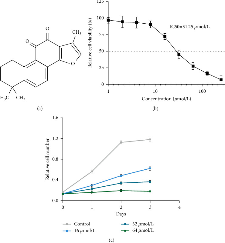
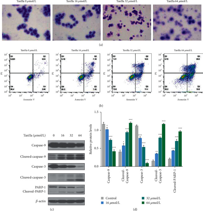
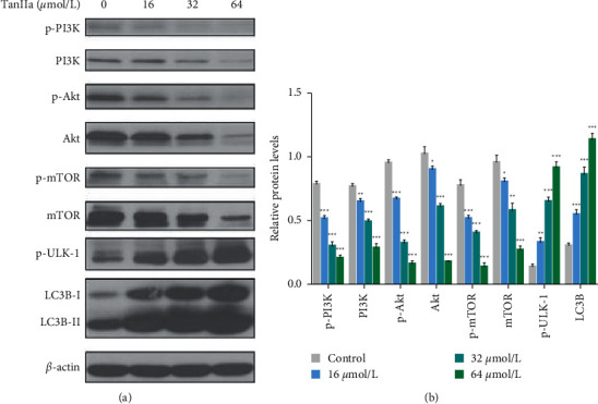
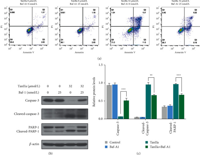
Tanshinone IIa (TanIIa), an ingredient of Radix Salviae Miltiorrhizae, has an anticancer effect on various solid tumors with high efficiency and low toxicity. Nonetheless, the underlying role of TanIIa in acute promyelocytic leukemia (APL) remains unclear. Here, we revealed that TanIIa drastically inhibited NB4 cell viability with an IC50 value of 31.25 μmol/L. Using flow cytometry apoptosis assay, we identified that TanIIa dose-dependently exacerbated NB4 cell apoptosis. Mechanistically, TanIIa upregulated apoptotic factor levels, namely, cleaved-caspase 9, cleaved-caspase 3, and cleaved-PARP-1. Moreover, we noticed that TanIIa dose-dependently suppressed the PI3K/Akt/mTOR axis. This axis not only functions as an essential antiapoptotic modulator but also serves as a suppressant regulator of autophagy. Correspondingly, we detected the levels of autophagic marker, namely, LC3B, which were increased after the TanIIa treatment. Furthermore, the autophagy inhibitor Baf-A1 could effectively reverse the TanIIa-induced apoptosis, manifesting that TanIIa eliminated NB4 cells in an autophagy-dependent manner. In conclusion, tanshinone IIa exerts anti-APL effects through triggering autophagy and apoptosis in NB4 cells.


Conflict of interest statement

The authors declare no conflicts of interest.

Figures

Figure 1
Figure 1
TanIIa inhibited NB4 viability and proliferation. (a) The chemical structure of TanIIa. (b) Cell viability assay: relative cell viability analyzed by CCK-8 in NB4 cells treated with different TanIIa doses, namely, 0, 1, 2, 4, 8, 16, 32, 64, 128, and 256 µmol/L for 24 h. Data were expressed as the mean ± SD of three independent experiments. (c) Cell proliferation assay: the relative cell number analyzed by CCK-8 in NB4 cells treated with TanIIa for 0–72 h. Data were expressed as the mean ± SD of three independent experiments.
Figure 2
Figure 2
TanIIa-induced NB4 apoptosis. (a) Giemsa staining of NB4 cells treated with TanIIa for 24 h. (b) Annexin V/PI staining of NB4 cells treated with TanIIa for 24 h. (c) The western blotting of apoptotic proteins in NB4 cells treated with TanIIa for 24 h. (d) The relative quantification of apoptotic proteins in NB4 cells treated with TanIIa for 24 h. Data were expressed as the mean ± SD of three independent experiments; P < 0.05,  ∗∗P < 0.01,  and ∗∗∗P < 0.001, compared with the 0 μmol/L groups.
Figure 3
Figure 3
TanIIa suppressed the PI3K/Akt/mTOR axis and activated autophagy in NB4 cells. (a) The western blotting of autophagic proteins in NB4 cells treated with TanIIa for 24 h. (b) The relative quantification of autophagic proteins in NB4 cells treated with TanIIa. Data were expressed as the mean ± SD of three independent experiments,  P < 0.05, ∗∗P < 0.01,  and  ∗∗∗P < 0.001, compared with the 0 μmol/L groups.
Figure 4
Figure 4
Blocking autophagy reduced the anti-APL effect of TanIIa. (a) The Annexin V/PI staining of NB4 cells treated with TanIIa or Baf-A1 or both for 24 h. (b) The western blotting of autophagic proteins in NB4 cells treated with TanIIa or Baf-A1 or both for 24 h. (c) The relative quantification of autophagic proteins in NB4 cells treated with TanIIa or Baf-A1 or both for 24 h. Data were expressed as the mean ± SD of three independent experiments,  ∗∗P < 0.01,  and  ∗∗∗P < 0.001, compared with the 0 μmol/L groups.

Similar articles

Cited by

References

    1. Park J. H., Qiao B., Panageas K. S., et al. Early death rate in acute promyelocytic leukemia remains high despite all-trans retinoic acid. Blood . 2011;118(5):1248–1254. doi: 10.1182/blood-2011-04-346437. - DOI - PMC - PubMed
    1. Zhu H., Hu J., Chen L., et al. The 12-year follow-up of survival, chronic adverse effects, and retention of arsenic in patients with acute promyelocytic leukemia. Blood . 2016;128(11):1525–1528. doi: 10.1182/blood-2016-02-699439. - DOI - PubMed
    1. Watts J. M., Tallman M. S. Acute promyelocytic leukemia: what is the new standard of care? Blood Reviews . 2014;28(5):205–212. doi: 10.1016/j.blre.2014.07.001. - DOI - PubMed
    1. Stahl M., Tallman M. S. Acute promyelocytic leukemia (APL): remaining challenges towards a cure for all. Leukemia and Lymphoma . 2019;60(13):3107–3115. doi: 10.1080/10428194.2019.1613540. - DOI - PMC - PubMed
    1. Shigeno K., Naito K., Sahara N., et al. Arsenic trioxide therapy in relapsed or refractory Japanese patients with acute promyelocytic leukemia: updated outcomes of the phase II study and postremission therapies. International Journal of Hematology . 2005;82(3):224–229. doi: 10.1532/ijh97.05044. - DOI - PubMed