

Exploring the Extracellular Vesicle MicroRNA Expression Repertoire in Patients with Rheumatoid Arthritis and Ankylosing Spondylitis Treated with TNF Inhibitors
- PMID: 34691284
- PMCID: PMC8529175
- DOI: 10.1155/2021/2924935
Exploring the Extracellular Vesicle MicroRNA Expression Repertoire in Patients with Rheumatoid Arthritis and Ankylosing Spondylitis Treated with TNF Inhibitors
Abstract
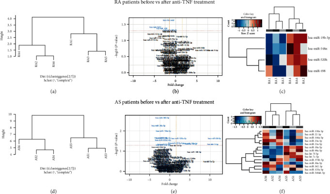
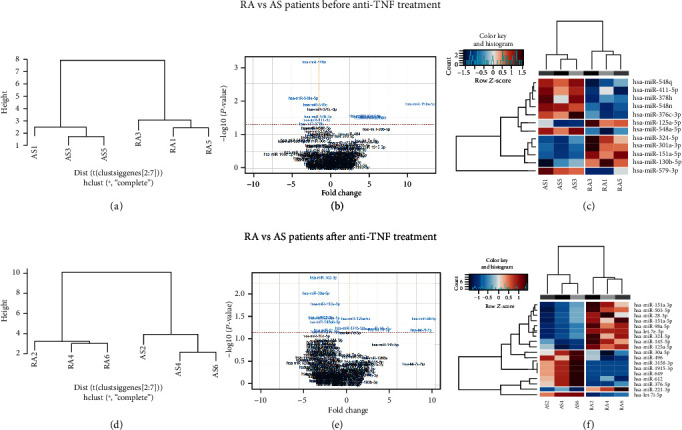
Rheumatoid arthritis (RA) and ankylosing spondylitis (AS) belong to the most common inflammatory rheumatic diseases. MicroRNAs (miRNAs) are small 18-22 RNA molecules that function as posttranscriptional regulators. They are abundantly present within extracellular vesicles (EVs), small intercellular communication vesicles that can be found in bodily fluids and that have key functions in pathological and physiological pathways. Recently, EVs have gained much interest because of their diagnostic and therapeutic potential. Using NanoString profiling technology, the miRNA repertoire of serum EVs was determined and compared in RA and AS patients before and after anti-TNF therapy to assess its potential use as a diagnostic and prognostic biomarker. Furthermore, possible functional effects of those miRNAs that were characterized by the most significant expression changes were evaluated using in silico prediction algorithms. The analysis revealed a unique profile of differentially expressed miRNAs in RA and AS patient serum EVs. We identified 12 miRNAs whose expression profiles enabled differentiation between RA and AS patients before induction of anti-TNF treatment, as well as 4 and 14 miRNAs whose repertoires were significantly changed during the treatment in RA and AS patients, respectively. In conclusion, our findings suggest that extracellular vesicle miRNAs could be used as potential biomarkers associated with RA and AS response to biological treatment.
Copyright © 2021 Joanna Wielińska et al.
Conflict of interest statement
The authors declare no conflict of interest.
Figures
Similar articles
-
IL-33 Gene Polymorphisms as Potential Biomarkers of Disease Susceptibility and Response to TNF Inhibitors in Rheumatoid Arthritis, Ankylosing Spondylitis, and Psoriatic Arthritis Patients.Front Immunol. 2021 Jun 11;12:631603. doi: 10.3389/fimmu.2021.631603. eCollection 2021. Front Immunol. 2021. PMID: 34177886 Free PMC article.
-
Changes in MiRNA-5196 Expression as a Potential Biomarker of Anti-TNF-α Therapy in Rheumatoid Arthritis and Ankylosing Spondylitis Patients.Arch Immunol Ther Exp (Warsz). 2018 Oct;66(5):389-397. doi: 10.1007/s00005-018-0513-y. Epub 2018 May 9. Arch Immunol Ther Exp (Warsz). 2018. PMID: 29744553 Free PMC article.
-
miRNAs as potential biomarkers of treatment outcome in rheumatoid arthritis and ankylosing spondylitis.Pharmacogenomics. 2021 Apr;22(5):291-301. doi: 10.2217/pgs-2020-0148. Epub 2021 Mar 26. Pharmacogenomics. 2021. PMID: 33769067 Review.
-
Extracellular Vesicles in Synovial Fluid from Rheumatoid Arthritis Patients Contain miRNAs with Capacity to Modulate Inflammation.Int J Mol Sci. 2021 May 6;22(9):4910. doi: 10.3390/ijms22094910. Int J Mol Sci. 2021. PMID: 34066338 Free PMC article.
-
Golimumab: a new anti-TNF-alpha agent for rheumatoid arthritis, psoriatic arthritis and ankylosing spondylitis.Expert Rev Clin Immunol. 2010 Sep;6(5):721-33. doi: 10.1586/eci.10.49. Expert Rev Clin Immunol. 2010. PMID: 20828280 Review.
Cited by
-
Genetic variability of three common NK and γδ T cell receptor genes (FCγ3R, NCR3, and DNAM-1) and their role in Polish patients with rheumatoid arthritis and ankylosing spondylitis.Immunol Res. 2024 Aug;72(4):614-625. doi: 10.1007/s12026-024-09488-3. Epub 2024 May 7. Immunol Res. 2024. PMID: 38714580 Free PMC article.
-
Identifying novel biomarkers for ankylosing spondylitis through proteomic profiling of serum-derived extracellular vesicles.Clin Exp Med. 2025 Jul 1;25(1):227. doi: 10.1007/s10238-025-01718-8. Clin Exp Med. 2025. PMID: 40591022 Free PMC article.
-
Year in Review: Novel Insights in the Pathogenesis of Spondyloarthritis - SPARTAN 2024 Annual Meeting Proceedings.Curr Rheumatol Rep. 2024 Dec 28;27(1):9. doi: 10.1007/s11926-024-01176-3. Curr Rheumatol Rep. 2024. PMID: 39731620 Review.
-
Updates on the Pathophysiology and Therapeutic Potential of Extracellular Vesicles with Focus on Exosomes in Rheumatoid Arthritis.J Inflamm Res. 2024 Jul 19;17:4811-4826. doi: 10.2147/JIR.S465653. eCollection 2024. J Inflamm Res. 2024. PMID: 39051053 Free PMC article. Review.
-
Biomarkers (mRNAs and non-coding RNAs) for the diagnosis and prognosis of rheumatoid arthritis.Front Immunol. 2023 Feb 1;14:1087925. doi: 10.3389/fimmu.2023.1087925. eCollection 2023. Front Immunol. 2023. PMID: 36817438 Free PMC article. Review.
References
MeSH terms
Substances
LinkOut - more resources
Full Text Sources
Medical
Research Materials
