. 2021 Oct 14:2021:5527137.
doi: 10.1155/2021/5527137. eCollection 2021.

Rosiglitazone Suppresses Renal Crystal Deposition by Ameliorating Tubular Injury Resulted from Oxidative Stress and Inflammatory Response via Promoting the Nrf2/HO-1 Pathway and Shifting Macrophage Polarization

Affiliations

Rosiglitazone Suppresses Renal Crystal Deposition by Ameliorating Tubular Injury Resulted from Oxidative Stress and Inflammatory Response via Promoting the Nrf2/HO-1 Pathway and Shifting Macrophage Polarization

Hongyan Lu et al. Oxid Med Cell Longev. .

Abstract

Oxidative stress and inflammatory response are closely related to nephrolithiasis. This study is aimed at exploring whether rosiglitazone (ROSI), a regulator of macrophage (Mp) polarization, could reduce renal calcium oxalate (CaOx) deposition by ameliorating oxidative stress and inflammatory response. Male C57 mice were equally and randomly divided into 7 groups. Kidney sections were collected on day 5 or day 8 after treatment. Pizzolato staining and polarized light optical microscopy were used to detect crystal deposition. PAS staining and TUNEL assay were performed to assess the tubular injury and cell apoptosis, respectively. Gene expression was assessed by immunohistochemistry, immunofluorescence, ELISA, qRT-PCR, and Western blot. The reactive oxygen species (ROS) level was assessed using a fluorescence microplate and fluorescence microscope. Hydrogen peroxide (H2O2), malonaldehyde (MDA), and glutathione (GSH) were evaluated to determine oxidative stress. Lactic dehydrogenase (LDH) activity was examined to detect cell injury. Adhesion of CaOx monohydrate (COM) crystals to HK-2 cells was detected by crystal adhesion assay. HK-2 cell death or renal macrophage polarization was assessed by flow cytometry. In vivo, renal crystal deposition, tubular injury, crystal adhesion, cell apoptosis, oxidative stress, and inflammatory response were significantly increased in the 7-day glyoxylic acid- (Gly-) treated group but were decreased in the ROSI-treated groups, especially in the groups pretreated with ROSI. Moreover, ROSI significantly reduced renal Mp aggregation and M1Mp polarization but significantly enhanced renal M2Mp polarization. In vitro, ROSI significantly suppressed renal injury, apoptosis, and crystal adhesion of HK-2 cells and markedly shifted COM-stimulated M1Mps to M2Mps, presenting an anti-inflammatory effect. Furthermore, ROSI significantly suppressed oxidative stress by promoting the Nrf2/HO-1 pathway in HK-2 cells. These findings indicate that ROSI could ameliorate renal tubular injury that resulted from oxidative stress and inflammatory response by suppressing M1Mp polarization and promoting M2Mp polarization. Therefore, ROSI is a potential therapeutic and preventive drug for CaOx nephrolithiasis.


Conflict of interest statement

The authors declare no conflicts of interest regarding the publication of this paper.

Figures

Figure 1
Figure 1
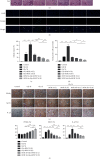
ROSI decreased the deposition of renal CaOx crystal in the mouse model. (a) Polarized light optical microscopy (arrows). ×20 magnification. (b) Pizzolato staining. Pizzolato staining indicates CaOx crystals (arrows). Scale bar = 50 μm. (c) The proportion of the crystal deposition area in the kidney and the proportion of crystal deposition areas in the corticomedullary border. (d) Immunohistochemical distribution of crystal-related gene OPN and crystal adhesion-related gene CD44. Scale bar = 50 μm. (e) The proportion of the IHC-positive area. Gly: glyoxylic acid; ROSI: rosiglitazone; CaOx: calcium oxalate. p < 0.05; ∗∗p < 0.01.
Figure 2
Figure 2
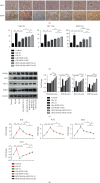
ROSI decreased renal tubular injury, cell apoptosis, and proinflammatory response in the mouse model. (a) PAS staining. PAS staining denotes tubular injury (arrows). Scale bar = 50 μm. (b) Cell apoptosis in the kidneys (arrows). Scale bar = 50 μm. (c) The percentage of damaged tubules displayed in PAS staining. (d) The mean number of apoptotic cells per high-power field (×400; n = 10 fields per section) in the TUNEL assay. (e) Immunohistochemical distribution of genetic expression of PPARγ, Mps-related molecule MCP1, and proinflammatory cytokine IL-1β. Scale bar = 50 μm. (f) The proportion of the IHC-positive area. Gly: glyoxylic acid; ROSI: rosiglitazone; PAS: periodic acid–Schiff; IL-1β: interleukin-1β; MCP1: monocyte chemotactic protein-1; PPARγ: peroxisome proliferator-activated receptor γ; IHC: immunohistochemistry. p < 0.05; ∗∗p < 0.01.
Figure 3
Figure 3
ROSI inhibited renal oxidative stress in the mouse model. (a) Immunohistochemical distribution of gene expression of Nrf2, HO-1, and SOD1 in the kidneys (arrows). Scale bar = 50 μm. (b) The proportion of the IHC-positive area. (c) The expression of PPARγ, Nrf2, HO-1, and SOD1 in kidney tissues by Western blotting. (d) ROS, H2O2, MDA, and GSH levels in mouse renal tissues. Gly: glyoxylic acid; ROSI: rosiglitazone. p < 0.05; ∗∗p < 0.01.
Figure 4
Figure 4
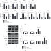
ROSI suppressed CaOx-induced oxidative stress injury and promoted the Nrf2/HO-1 pathway in HK-2 cell. (a) Cellular LDH levels in HK-2 cells. (b) HK-2 cell death by flow cytometry. (c) mRNA expression levels of Nrf2, HO-1, and SOD1 in HK-2 cells by qRT-PCR. (d) The expression of PPARγ, Nrf2, HO-1, and SOD1 in HK-2 cells by Western blot. (e) Detection of intracellular ROS levels by a fluorescence microscope. (f) H2O2, MDA, and GSH levels in HK-2 cells. COM: calcium oxalate monohydrate; ROSI: rosiglitazone. p < 0.05; ∗∗p < 0.01.
Figure 5
Figure 5
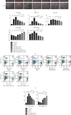
ROSI decreased renal macrophage recruitment and M1Mp polarization and promoted renal M2Mp polarization in the mouse model. (a) Immunohistochemical distribution of genetic expression of PPARγ, macrophage-related molecules, and proinflammatory cytokines (arrows). Scale bar = 50 μm. (b) The proportion of the IHC-positive area and the ratio of Arg1/F4/80 and iNOS/F4/80. (c) Flow cytometric detection of Mps in kidney tissues. (d) The proportion of M1Mps and M2Mps in kidney tissues. Gly: glyoxylic acid; ROSI: rosiglitazone; PPARγ: peroxisome proliferator-activated receptor γ. p < 0.05; ∗∗p < 0.01.
Figure 6
Figure 6
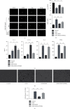
ROSI declined COM-stimulated M1Mp polarization and crystal adhesion and promoted M2Mp polarization in vitro. (a) Fluorescence immunohistochemical distribution of Arg1 (green) and iNOS (red) in THP-1 cells with the stimulus of COM or ROSI (1 μM) or GW9662 (10 μM) (arrows). (b) Evaluation of mean fluorescence intensity (MFI) of Arg1 and iNOS. (c) ELISA. The expression levels of IL-4, IL-10, IL-6, and TNF-α. (d) Crystal adhesion assay. COM crystal adhesion to HK-2 cells was observed (arrows). COM: calcium oxalate monohydrate; ROSI: rosiglitazone; iNOS: induced nitric oxide synthase; TNF-α: tumor necrosis factor α. p < 0.05; ∗∗p < 0.01.
Figure 7
Figure 7
Genetic expression of macrophage-related molecules, proinflammatory cytokines, and anti-inflammatory cytokines. (a) mRNA levels in THP-1 cells stimulated with COM or ROSI (1 μM) or GW9662 (10 μM) by qRT-PCR. (b) Genetic expression determined by Western blot. COM: calcium oxalate monohydrate; ROSI: rosiglitazone. p < 0.05; ∗∗p < 0.01.
Figure 8
Figure 8
Mechanisms by which ROSI suppresses CaOx crystal deposition. ROSI: rosiglitazone; PPARγ: peroxisome proliferator-activated receptor γ.

References

    1. Neisius A., Preminger G. M. Stones in 2012: epidemiology, prevention and redefining therapeutic standards . - PubMed
    1. Zisman A. L., Evan A. P., Coe F. L., Worcester E. M. Do kidney stone formers have a kidney disease? Kidney International . 2015;88(6):1240–1249. doi: 10.1038/ki.2015.254. - DOI - PMC - PubMed
    1. Antonelli J. A., Maalouf N. M., Pearle M. S., Lotan Y. Use of the National Health and Nutrition Examination Survey to calculate the impact of obesity and diabetes on cost and prevalence of urolithiasis in 2030. European Urology . 2014;66(4):724–729. doi: 10.1016/j.eururo.2014.06.036. - DOI - PMC - PubMed
    1. Khan S. R., Byer K. J., Thamilselvan S., et al. Crystal-cell interaction and apoptosis in oxalate-associated injury of renal epithelial cells. Journal of the American Society of Nephrology . 1999;10(Supplement 14):S457–S463. - PubMed
    1. Taguchi K., Okada A., Hamamoto S., et al. Proinflammatory and metabolic changes facilitate renal crystal deposition in an obese mouse model of metabolic syndrome. Journal of Urology . 2015;194(6):1787–1796. doi: 10.1016/j.juro.2015.07.083. - DOI - PubMed

Supplementary concepts