

Retroperitoneal shwannoma: A case report
- PMID: 34691409
- PMCID: PMC8519761
- DOI: 10.1016/j.amsu.2021.102785
Retroperitoneal shwannoma: A case report
Abstract
Introduction: Schwannomas are tumors that arise from Schwann cells of the peripheral nerve sheath and rarely occur in the retroperitoneum (3% of all schwannomas). Patients are usually asymptomatic or have nonspecific symptoms, making accurate preoperative diagnosis difficult. Schwannomas are usually benign, but infrequently undergo malignant transformation. Herein, we report a case of retroperitoneal schwannoma and review the relevant literature.
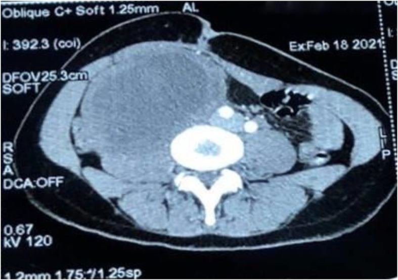
Presentation of case: A 25-year-old woman presented to our department with a 2-year history of abdominal pain that was localized in the right flank without radiation, constipation/diarrhea or externalized digestive hemorrhage. On physical examination, we found a painless palpable mass in the right hypochondrium extending to the right iliac fossa, measuring approximately 10 cm. The MRI and CT scan showed the presence of a large intra-abdominal oval formation in the right para-umbilical region. It was well limited, measuring 110*69mm with discrete irregular contours, thickened wall and heterogeneous content mostly fluid. They also showed the presence of a cystic formation in the right ovary measuring 84*52mm and extending over 76mm. The procedure consisted of resection of the retroperitoneal solid cystic mass, right ovariectomy and drainage of the right parietal-colic gutter by Salem sump tube. A laparotomy with a median incision above and below the umbilicus was performed. After the resection, the specimens were sent for anatomopathological examination which concluded that the retroperitoneal mass was a schwannoma and the ovarian mass was a serous cystadenoma.
Discussion: Retroperitoneal schwannomas are rare tumors and a pre-operative diagnosis is often difficult. The diagnosis is most often fortuitous and late, given the latency of the tumor's evolution, and the definitive diagnosis is based on histopathologic examination. Herein we presented a case of retroperitoneal schwannoma and studied the features of this phenomenon on the basis of the literature.
Conclusion: Retroperitoneal schwannomas are rare. The diagnosis is often late at the stage of a large tumor. Radiologic findings are usually nondiagnostic. The treatment of choice is complete surgical excision. Prognosis is good but because of the risk of recurrence and malignant transformation, further follow-up is necessary.
Keywords: Case report; Retroperitoneal; Schwann cells; Schwannomas.
© 2021 Published by Elsevier Ltd on behalf of IJS Publishing Group Ltd.
Conflict of interest statement
The authors report no declarations of interest.
Figures


References
-
- Agha R.A., Franchi T., Sohrabi C., Mathew G., Kerwan A., Thoma A. The SCARE 2020 guideline: updating consensus Surgical CAse REport (SCARE) guidelines. Int. J. Surg. 2020;84:226–230. - PubMed
-
- Ben Moualli S., Hajri M., Ben Amna M., Kolsi K., Chebil M., Ben Jilani S. Le schwannome rétropéritonéal. À propos d’un cas. Ann. Urol. 2001;35(5):270–272. - PubMed
Publication types
LinkOut - more resources
Full Text Sources
Research Materials
