. 2020 Jun 10;8(1):nwaa122.
doi: 10.1093/nsr/nwaa122. eCollection 2021 Jan.

Biodegradable magnesium alloy with eddy thermal effect for effective and accurate magnetic hyperthermia ablation of tumors

Affiliations

Biodegradable magnesium alloy with eddy thermal effect for effective and accurate magnetic hyperthermia ablation of tumors

Nailin Yang et al. Natl Sci Rev. .

Abstract

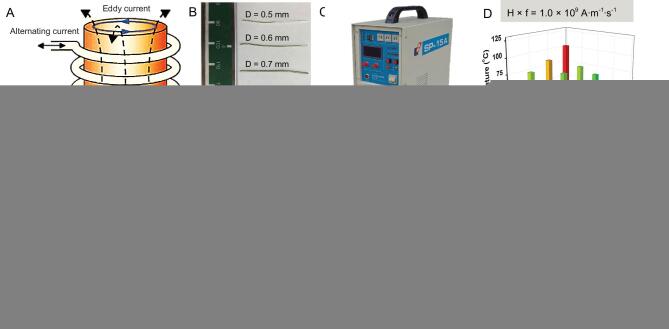
Magnetic hyperthermia therapy (MHT) is able to ablate tumors using an alternating magnetic field (AMF) to heat up magnetocaloric agents (e.g. magnetic nanoparticles) administered into the tumors. For clinical applications, there is still a demand to find new magnetocaloric agents with strong AMF-induced heating performance and excellent biocompatibility. As a kind of biocompatible and biodegradable material, magnesium (Mg) and its alloys have been extensively used in the clinic as an implant metal. Herein, we discovered that the eddy thermal effect of the magnesium alloy (MgA) could be employed for MHT to effectively ablate tumors. Under low-field-intensity AMFs, MgA rods could be rapidly heated, resulting in a temperature increase in nearby tissues. Such AMF-induced eddy thermal heating of MgA could not only be used to kill tumor cells in vitro, but also be employed for effective and accurate ablation of tumors in vivo. In addition to killing tumors in mice, we further demonstrated that VX2 tumors of much larger sizes growing in rabbits after implantation of MgA rods could also be eliminated after exposure to an AMF, illustrating the ability of MgA-based MHT to kill large-sized tumors. Moreover, the implanted MgA rods showed excellent biocompatibility and ∼20% of their mass was degraded within three months. Our work thus discovered for the first time that non-magnetic biodegradable MgA, an extensively used implant metal in clinic, could be used for effective magnetic thermal ablation of tumors under a low-field-intensity AMF. Such a strategy could be readily translated into clinical use.

Keywords: alternating magnetic field; biodegradation; eddy thermal effect; hyperthermia tumor ablation; magnesium alloy.


Figures

Scheme 1.
Scheme 1.
Schematic illustration of MHT based on the eddy thermal effect of the biodegradable MgA rods.
Figure 1.
Figure 1.
AMF-induced eddy thermal effect of MgA rods. (A) Schematic illustration to show AMF-induced eddy thermal heating of MgA rods. Photograph of MgA rods with different diameters (D: 0.50 ± 0.01, 0.60 ± 0.01, 0.70 ± 0.01, 0.80 ± 0.01 and 0.90 ± 0.01 mm) (B) and the high-frequency AMF induction equipment (C). (D–H) The temperature increase data of MgA rods with different lengths (2.0 ± 0.1, 4.0 ± 0.1, 6.0 ± 0.1 and 8.0 ± 0.1 mm) or different diameters (0.50 ± 0.01, 0.60 ± 0.01, 0.70 ± 0.01, 0.80 ± 0.01 and 0.90 ± 0.01 mm) under AMF with different field intensities.
Figure 2.
Figure 2.
Effective killing ranges based on heating of pork tissues with MgA rods under AMFs. (A) Thermal imaging of MgA rod (D = 0.7 mm, L = 4.0 mm) under AMF with different field intensities (Happl × fappl = M × 109 A · m−1 · s−1; M = 1.5, 1.75, 2.0, 2.25 and 2.5). (B) Schematic illustration of the effective killing range simulation. (C) The effective killing range of MgA rods under AMF with different field intensities based on the heat diffusion ranges in pork tissues with the temperature above 50°C. a: length; b: width.
Figure 3.
Figure 3.
In vitro MHT with MgA rods. (A) Ion concentrations in RPMI cell culture medium immersed with MgA rods with different periods of time. (B) The viabilities of L929 cells, 4T1 cells and VX2 cells after 24 h of exposure to MgA-treated cell culture medium. (C) Relative viabilities of 4T1 cells and VX2 cells after MHT treated by MgA rods. (D) Confocal fluorescence images of VX2 cells stained with Calcein AM (AM, green, live cells) and propidium iodide (PI, red, dead cells) after various treatments. The temperature of treatment was controlled at ∼42°C (Happl × fappl = 2.0 × 10⁹ A · m−1 · s−1), ∼48°C (Happl × fappl = 2.5 × 10⁹ A · m−1 · s−1) and ∼54°C (Happl × fappl = 3.0 × 10⁹ A · m−1 · s−1), respectively.
Figure 4.
Figure 4.
In vivo MHT treatment of 4T1 mouse tumors with MgA rods. (A) Schematic illustration of MHT with MgA rods to ablate tumors in mice. Infrared thermal images (B) and temperature change curves (C) of tumors in mice with or without implantation of MgA rods under AMF. (D) The growth curves of tumors after various treatments. (E) H&E stained tumor slices collected from different treatment groups.
Figure 5.
Figure 5.
In vivo MHT treatment of VX2 rabbit tumors with MgA rods. (A) Schematic illustration of rabbit VX2 tumor model. Infrared thermal images (B) and temperature change curves (C) of tumors in rabbits with or without implantation of MgA rods under AMF. (D) The growth curves of tumors after various treatments. (E) Survival rates of tumor-bearing rabbits post various treatments. The tumor in one out of three rabbits was not completely eliminated by MgA-based MHT and later showed re-growth.
Figure 6.
Figure 6.
Biodegradation behaviors of MgA rods. (A) Scanning electron microscope (SEM) images to show the surfaces on MgA rods after in vivo implantation into mice for different periods of time. (B) The time-dependent mass loss of MgA rods after in vivo implantation into mice. (C) The Mg levels in major organs of mice after being implanted with MgA rods for different numbers of days. The Mg contents were measured by ICP-OES. No notable increase of Mg2+ levels was found in organs of implanted mice compared to the blank control. (D, E) The mass loss of MgA rods of two different dimensions (D: 0.7 mm × 4 mm; E: 1.0 mm × 4 mm) after being immersed in different solutions for different periods of time. (F) The XRD patterns of MgA after being immersed in PBS for 90 days.

References

    1. Bañobre-López M, Teijeiro A, Rivas J. Magnetic nanoparticle-based hyperthermia for cancer treatment. Rep Pract Oncol Radiother 2013; 18: 397–400. - PMC - PubMed
    1. Kumar CS, Mohammad F. Magnetic nanomaterials for hyperthermia-based therapy and controlled drug delivery. Adv Drug Delivery Rev 2011; 63: 789–808. - PMC - PubMed
    1. Dutz S, Hergt R. Magnetic nanoparticle heating and heat transfer on a microscale: basic principles, realities and physical limitations of hyperthermia for tumour therapy. Int J Hyperthermia 2013; 29: 790–800. - PubMed
    1. Hilger I, Hiergeist R, Hergt Ret al. . Thermal ablation of tumors using magnetic nanoparticles: an in vivo feasibility study. Invest Radiol 2002; 37: 580–6. - PubMed
    1. Tishin A, Spichkin Y, Zverev Vet al. . A review and new perspectives for the magnetocaloric effect: new materials and local heating and cooling inside the human body. Int J Refrig 2016; 68: 177–86.