Diagnostic Ability of High-definition Imaging Using Ultraslim Endoscopes in Early Gastric Cancer
- PMID: 34691809
- PMCID: PMC8505118
- DOI: 10.5230/jgc.2021.21.e23
Diagnostic Ability of High-definition Imaging Using Ultraslim Endoscopes in Early Gastric Cancer
Abstract
Purpose: It is unclear whether high-definition (HD) imaging improves visibility and diagnostic ability in early gastric cancer (EGC) compared with standard-definition (SD) imaging. We aimed to compare the diagnostic performance and visibility scores of HD and SD ultraslim endoscopes in EGC.
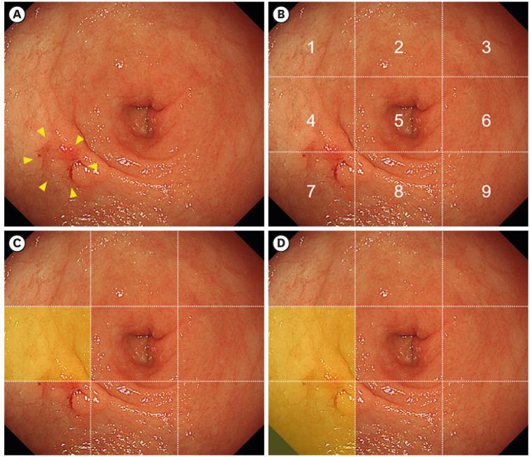
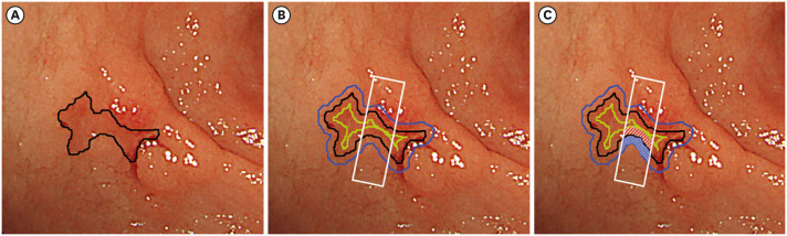
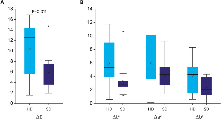
Materials and methods: We used HD and SD ultraslim endoscopes to obtain 60 images with similar compositions of gastric environments. Of the 60 images, 30 showed EGC (15 images for each modality) and 30 showed no EGC (15 images for each modality). Seventeen endoscopists evaluated the presence and location of the lesions in each image. Diagnostic ability was compared between modalities. The color difference between a lesion and the surrounding mucosa (ΔE) was measured and compared between the modalities.
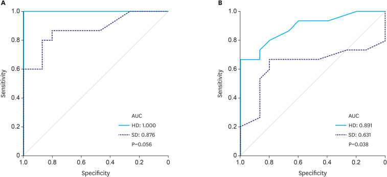
Results: The ability of HD to detect EGC was significantly higher than that of SD (accuracy: 80.8% vs. 71.6%, P=0.017; sensitivity: 94.9% vs. 76.5%, P<0.001; positive predictive value, 76.2% vs. 55.3%, P<0.001; and negative predictive value (NPV), 94.1% vs. 73.5%, P<0.001). The ability of HD to determine the horizontal extent of EGC was significantly higher than that of SD (accuracy: 71.0% vs. 57.8%, P=0.004; sensitivity: 75.3% vs. 49.0%, P<0.001; NPV, 72.9% vs. 55.9%, P<0.001; and area under the curve: 0.891 vs. 0.631, P=0.038). The mean ΔE was significantly higher for HD than for SD (10.3 vs. 5.9, P=0.011).
Conclusions: The HD ultraslim endoscope showed a higher diagnostic performance in EGC than the SD endoscope because it provided good color contrast.
Keywords: Diagnosis; Diagnostic imaging; Endoscopy; Gastric cancer; Screening.
Copyright © 2021. Korean Gastric Cancer Association.
Conflict of interest statement
Conflict of Interest: Takuji Gotoda received an honorarium from Olympus Corporation.
Figures
Similar articles
-
Visibility of early gastric cancers by texture and color enhancement imaging using a high-definition ultrathin transnasal endoscope.Sci Rep. 2023 Feb 3;13(1):1994. doi: 10.1038/s41598-023-29284-7. Sci Rep. 2023. PMID: 36737509 Free PMC article.
-
Magnifying narrow-band imaging endoscopy is superior in diagnosis of early gastric cancer.World J Gastroenterol. 2015 Aug 14;21(30):9156-62. doi: 10.3748/wjg.v21.i30.9156. World J Gastroenterol. 2015. PMID: 26290643 Free PMC article.
-
Visibility of early gastric cancer in texture and color enhancement imaging.DEN Open. 2021 Aug 24;2(1):e46. doi: 10.1002/deo2.46. eCollection 2022 Apr. DEN Open. 2021. PMID: 35310718 Free PMC article.
-
Current Evidence and Future Perspective of Accuracy of Artificial Intelligence Application for Early Gastric Cancer Diagnosis With Endoscopy: A Systematic and Meta-Analysis.Front Med (Lausanne). 2021 Mar 15;8:629080. doi: 10.3389/fmed.2021.629080. eCollection 2021. Front Med (Lausanne). 2021. PMID: 33791323 Free PMC article.
-
Diagnostic Ability of Magnifying Narrow-Band Imaging for the Extent of Early Gastric Cancer: A Systematic Review and Meta-Analysis.Gastroenterol Res Pract. 2021 Apr 23;2021:5543556. doi: 10.1155/2021/5543556. eCollection 2021. Gastroenterol Res Pract. 2021. PMID: 33986796 Free PMC article. Review.
Cited by
-
Visibility of early gastric cancers by texture and color enhancement imaging using a high-definition ultrathin transnasal endoscope.Sci Rep. 2023 Feb 3;13(1):1994. doi: 10.1038/s41598-023-29284-7. Sci Rep. 2023. PMID: 36737509 Free PMC article.
-
Diagnostic performance of deep-learning-based virtual chromoendoscopy in gastric neoplasms.Gastric Cancer. 2024 May;27(3):539-547. doi: 10.1007/s10120-024-01469-7. Epub 2024 Jan 19. Gastric Cancer. 2024. PMID: 38240891
References
-
- Bray F, Ferlay J, Soerjomataram I, Siegel RL, Torre LA, Jemal A. Global cancer statistics 2018: GLOBOCAN estimates of incidence and mortality worldwide for 36 cancers in 185 countries. CA Cancer J Clin. 2018;68:394–424. - PubMed
-
- Choi IJ, Lee JH, Kim YI, Kim CG, Cho SJ, Lee JY, et al. Long-term outcome comparison of endoscopic resection and surgery in early gastric cancer meeting the absolute indication for endoscopic resection. Gastrointest Endosc. 2015;81:333–341.e1. - PubMed
-
- Gu L, Khadaroo PA, Chen L, Li X, Zhu H, Zhong X, et al. Comparison of long-term outcomes of endoscopic submucosal dissection and surgery for early gastric cancer: a systematic review and meta-analysis. J Gastrointest Surg. 2019;23:1493–1501. - PubMed
-
- Jeon HK, Kim GH, Lee BE, Park DY, Song GA, Kim DH, et al. Long-term outcome of endoscopic submucosal dissection is comparable to that of surgery for early gastric cancer: a propensity-matched analysis. Gastric Cancer. 2018;21:133–143. - PubMed
LinkOut - more resources
Full Text Sources