. 2021 Aug 11;12(9):5499-5513.
doi: 10.1364/BOE.431397. eCollection 2021 Sep 1.

Dec-DISCO: decolorization DISCO clearing for seeing through the biological architectures of heme-rich organs

Affiliations

Dec-DISCO: decolorization DISCO clearing for seeing through the biological architectures of heme-rich organs

Jingtan Zhu et al. Biomed Opt Express. .

Abstract

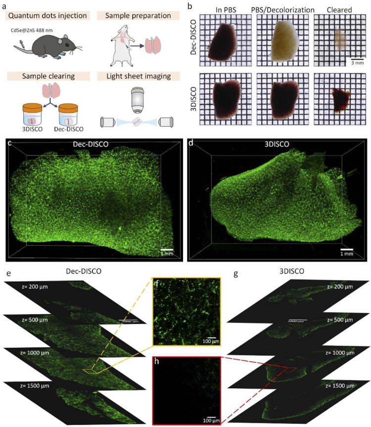
The tissue optical clearing technique plays an important role in three-dimensional (3D) visualization of large tissues. As a typical solvent-based clearing method, 3DISCO can achieve the highest level of tissue transparency with favorable clearing speed. However, 3DISCO cannot deal with the residual blood within tissues, leading to tissue brownness or redness after clearing, thus greatly influencing the tissue transparency and image quality due to the strong absorption of residual blood. To address this problem, we proposed an optimized clearing method by introducing CUBIC-L solution combined with 3DISCO for effective decolorization, termed Dec-DISCO (Decolorization DISCO). Dec-DISCO achieves better transparency than 3DISCO for various heme-rich tissues and performs enhanced fluorescence preservation capability. Dec-DISCO allows high-quality 3D imaging of fluorescently labeled heme-rich organs, as well as pathological tissue with severe hemorrhage. Dec-DISCO is expected to provide a powerful tool for histological analysis of kinds of heme-rich tissues in various medical conditions.


Conflict of interest statement

The authors declare no conflicts of interest.

Figures

Fig. 1.
Fig. 1.
Modifying 3DISCO by tissue decolorization. (a) Work flow for evaluating the decolorization abilities for chosen solutions. (b) Bright-field images of adult mouse spleens treated by different decolorization reagents. (c) Experimental pipeline for quantifying the fluorescence preservation abilities for different decolorization solutions. (d) Fluorescence images of mouse brain slices after decolorization by indicated solution and after 3DISCO clearing. All confocal images are MIPs of z stacks (50 µm thick) from the surface. (e) Comparison of fluorescence intensity of brain samples after decolorization by different reagents prior to clearing (n=6). (f) Comparison of fluorescence intensity of decolorized samples after 3DISCO clearing (n=6). All values are presented as the mean ± SD. Statistical signifcance in e and f (***, P < 0.001) was assessed by one-way ANOVA followed by the Bonferroni post hoc test.
Fig. 2.
Fig. 2.
Dec-DISCO allows high-performance clearing for heme-rich rodent tissues. (a) The entire pipeline for Dec-DISCO clearing. (b) Bright-field images of adult mouse kidneys cleared by 3DISCO and Dec-DISCO, respectively. (c) Transmittance curves of adult mouse kidneys cleared by 3DISCO and Dec-DISCO (n = 3). (d) Bright-field images of different rat organs cleared by 3DISCO and Dec-DISCO. All values are presented as the mean ± SD.
Fig. 3.
Fig. 3.
Dec-DISCO is applicable for high-quality imaging of neural structures in the mouse brain using LSFM. (a) The experimental work flow. (b) Bright-field images of mouse hemispheres cleared by Dec-DISCO and 3DISCO, respectively. (c) 3D reconstruction of neural structures in the mouse hemisphere cleared by Dec-DISCO using LSFM. (d) Cross-sections of the hemisphere at different imaging depths cleared by Dec-DISCO. (e) Cross-sections of the hemisphere at different imaging depths cleared by 3DISCO. (f) 3D reconstruction of neural structures in the mouse hemisphere cleared by 3DISCO. (g) Comparison of the high-magnification images in different brain regions after Dec-DISCO and 3DISCO clearing. (h) Quantification of the fluorescence level in different brain regions after Dec-DISCO and 3DISCO. The same imaging parameters were used for imaging the brain samples cleared by Dec-DISCO and 3DISCO (n=3). All values are presented as the mean ± SD. Statistical significance in h (***, P < 0.001) was assessed by an independent-sample t-test.
Fig. 4.
Fig. 4.
Dec-DISCO achieved complete 3D imaging with no blind area for heme-rich mouse lungs labeled by quantum dots. (a) Experimental design for clearing and imaging of mouse lungs labeled by quantum dots. (b) Bright-field images of labeled mouse lungs before and after clearing with Dec-DISCO and 3DISCO, respectively. (c) 3D reconstruction of cell populations labeled by quantum dots in the mouse lungs cleared by Dec-DISCO. (d) 3D reconstruction of cell populations labeled by quantum dots in the mouse lungs cleared by 3DISCO. (e) Cross-section images of the lungs in c at different imaging depths. (f) Magnification of boxed regions in e. (g) Cross-section images of the lungs in d at different imaging depths. (h) Magnification of boxed regions in g.
Fig. 5.
Fig. 5.
Dec-DISCO permits 3D visualization of neural disruptions in injured spinal cord with severe hemorrhage. (a) Experimental design for clearing and imaging of injured spinal cords from spinal cord injury model. (b) Bright-field images of injured spinal cords before and after clearing with Dec-DISCO and 3DISCO. (c) 3D imaging of neural structures of injured spinal cord cleared by Dec-DISCO. (d) 3D imaging of neural structures of injured spinal cord cleared by 3DISCO. (e, f) Visualization of neural information in normal (e) and injured area (f) cleared by Dec-DISCO. (g, h) Visualization of neural information in normal (g) and injured area (h) cleared by 3DISCO.

References

    1. Ueda H. R., Erturk A., Chung K., Gradinaru V., Chedotal A., Tomancak P., Keller P. J., “Tissue clearing and its applications in neuroscience,” Nat. Rev. Neurosci. 21(2), 61–79 (2020).10.1038/s41583-019-0250-1 - DOI - PMC - PubMed
    1. Zhao S., Todorov M. I., Cai R., Maskari R. A., Steinke H., Kemter E., Mai H., Rong Z., Warmer M., Stanic K., Schoppe O., Paetzold J. C., Gesierich B., Wong M. N., Huber T. B., Duering M., Bruns O. T., Menze B., Lipfert J., Puelles V. G., Wolf E., Bechmann I., Erturk A., “Cellular and Molecular Probing of Intact Human Organs,” Cell 180(4), 796–812.e19 (2020).10.1016/j.cell.2020.01.030 - DOI - PMC - PubMed
    1. Yu T., Qi Y., Gong H., Luo Q., Zhu D., “Optical clearing for multiscale biological tissues,” J. Biophotonics 11(2), e201700187 (2018).10.1002/jbio.201700187 - DOI - PubMed
    1. Costantini I., Cicchi R., Silvestri L., Vanzi F., Pavone F. S., “In-vivo and ex-vivo optical clearing methods for biological tissues: review,” Biomed. Opt. Express 10(10), 5251–5267 (2019).10.1364/BOE.10.005251 - DOI - PMC - PubMed
    1. Zhu D., Larin K. V., Luo Q., Tuchin V. V., “Recent progress in tissue optical clearing,” Laser Photonics Rev. 7(5), 732–757 (2013).10.1002/lpor.201200056 - DOI - PMC - PubMed

LinkOut - more resources