

Metastatic atypical renal tumour with metanephric characteristics treated with Sunitinib
- PMID: 34692420
- PMCID: PMC8517833
- DOI: 10.1016/j.eucr.2021.101880
Metastatic atypical renal tumour with metanephric characteristics treated with Sunitinib
Abstract
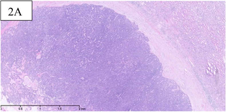
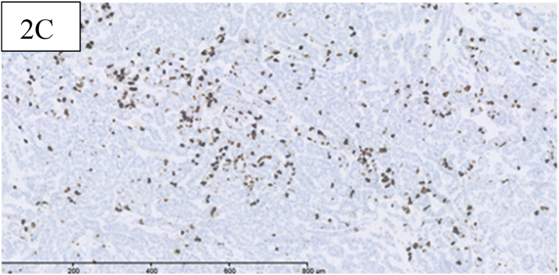
Metanephric Adenoma (MA) is a rare and unclassifiable renal tumour with sparse reported clinical and morphological features. Generally MA's have a benign course without recurrence after nephrectomy, however a few cases received oncological treatment due to malignant progression. We present a 42-year-old woman who years after an initial nephrectomy developed several processes and biopsy confirmed recurrence of MA. Sunitinib was given for only two weeks, as she developed side-effects and currently the patient undergoes control scans with only minimal growth of the processes. This is the first case of MA treated with Tyrosin-Kinase-Inhibitor.
Keywords: Metanephric adenoma; Oncology; Renal tumour; Tyrosine kinase inhibitor.
© 2021 The Authors. Published by Elsevier Inc.
Figures


References
-
- Davis C., Barton J., Sesterhenn I. Clinicopathological study of fifty patients. Am J Surg Pathol. 1995;19(10):1101–1114. - PubMed
-
- Pins M., Jones E., Martul V., Kamat B., Unlas J., Renshaw A. Metanephric adenoma- like tumor of the kidney. Report of 3 malignancies with emphasis on discriminating features. Arch Pathol Lab Med. 1999;123(5):415–420. - PubMed
-
- Brown J.A., Anderl K.L., Borell T.J., Qian J., Bostwick D.G., Jenkins R.B. Simultaneous chromosome 7 and 17 gain and sex chromosome loss provide evidence that renal metanephric adenoma is related to papillary renal cell carcinoma. J Urol. 1997;158:370–374. - PubMed
-
- Renshaw A.A., Freyer D.R., Hammers Y.A. Metastatic metanephric adenoma in a child. Am J Surg Pathol. 2000;24(4):570–574. - PubMed
Publication types
LinkOut - more resources
Full Text Sources
