

The Potential Roles of Glial Cells in the Neuropathogenesis of Cerebral Malaria
- PMID: 34692564
- PMCID: PMC8529055
- DOI: 10.3389/fcimb.2021.741370
The Potential Roles of Glial Cells in the Neuropathogenesis of Cerebral Malaria
Abstract
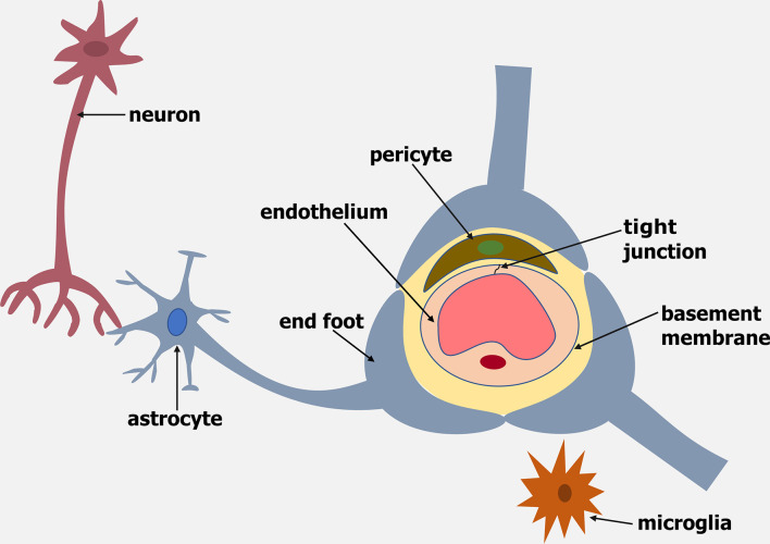
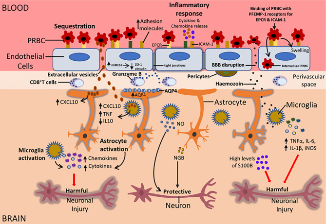
Cerebral malaria (CM) is a severe neurological complication of malaria caused by the Plasmodium falciparum parasite. It is one of the leading causes of death in children under 5 years of age in Sub-Saharan Africa. CM is associated with blood-brain barrier disruption and long-term neurological sequelae in survivors of CM. Despite the vast amount of research on cerebral malaria, the cause of neurological sequelae observed in CM patients is poorly understood. In this article, the potential roles of glial cells, astrocytes, and microglia, in cerebral malaria pathogenesis are reviewed. The possible mechanisms by which glial cells contribute to neurological damage in CM patients are also examined.
Keywords: Plasmodium; astrocytes; blood-brain barrier; cerebral malaria; glial cells; microglia.
Copyright © 2021 Andoh and Gyan.
Conflict of interest statement
The authors declare that the research was conducted in the absence of any commercial or financial relationships that could be construed as a potential conflict of interest.
Figures
Similar articles
-
Cerebral malaria induced by plasmodium falciparum: clinical features, pathogenesis, diagnosis, and treatment.Front Cell Infect Microbiol. 2022 Jul 25;12:939532. doi: 10.3389/fcimb.2022.939532. eCollection 2022. Front Cell Infect Microbiol. 2022. PMID: 35959375 Free PMC article. Review.
-
Uptake of parasite-derived vesicles by astrocytes and microglial phagocytosis of infected erythrocytes may drive neuroinflammation in cerebral malaria.Glia. 2017 Jan;65(1):75-92. doi: 10.1002/glia.23075. Epub 2016 Oct 3. Glia. 2017. PMID: 27696532
-
Pathophysiology and neurologic sequelae of cerebral malaria.Malar J. 2020 Jul 23;19(1):266. doi: 10.1186/s12936-020-03336-z. Malar J. 2020. PMID: 32703204 Free PMC article. Review.
-
Pattern and predictors of neurological morbidities among childhood cerebral malaria survivors in central Sudan.J Vector Borne Dis. 2015 Sep;52(3):239-44. J Vector Borne Dis. 2015. PMID: 26418655
-
Cerebral malaria: mechanisms of brain injury and strategies for improved neurocognitive outcome.Pediatr Res. 2010 Oct;68(4):267-74. doi: 10.1203/PDR.0b013e3181eee738. Pediatr Res. 2010. PMID: 20606600 Free PMC article. Review.
Cited by
-
Dynamic intravital imaging reveals reactive vessel-associated microglia play a protective role in cerebral malaria coagulopathy.Sci Rep. 2023 Nov 9;13(1):19526. doi: 10.1038/s41598-023-43208-5. Sci Rep. 2023. PMID: 37945689 Free PMC article.
-
The spatiotemporal transcriptional profiling of murine brain during cerebral malaria progression and after artemisinin treatment.Nat Commun. 2025 Feb 11;16(1):1540. doi: 10.1038/s41467-024-52223-7. Nat Commun. 2025. PMID: 39934099 Free PMC article.
-
Infections in the Developing Brain: The Role of the Neuro-Immune Axis.Front Neurol. 2022 Feb 17;13:805786. doi: 10.3389/fneur.2022.805786. eCollection 2022. Front Neurol. 2022. PMID: 35250814 Free PMC article. Review.
-
Neurons upregulate PD-L1 via IFN/STAT1/IRF1 to alleviate damage by CD8+ T cells in cerebral malaria.J Neuroinflammation. 2024 May 7;21(1):119. doi: 10.1186/s12974-024-03114-7. J Neuroinflammation. 2024. PMID: 38715061 Free PMC article.
-
Microglial Priming in Infections and Its Risk to Neurodegenerative Diseases.Front Cell Neurosci. 2022 Jun 15;16:878987. doi: 10.3389/fncel.2022.878987. eCollection 2022. Front Cell Neurosci. 2022. PMID: 35783096 Free PMC article. Review.
References
-
- Adams Y., Olsen R. W., Bengtsson A., Dalgaard N., Zdioruk M., Satpathi S., et al. . (2021). Plasmodium Falciparum Erythrocyte Membrane Protein 1 Variants Induce Cell Swelling and Disrupt the Blood–Brain Barrier in Cerebral Malaria. J. Exp. Med. 218, e20201266. doi: 10.1084/jem.20201266 - DOI - PMC - PubMed
Publication types
MeSH terms
LinkOut - more resources
Full Text Sources
Medical