Review
. 2021 Oct 8:9:751079.
doi: 10.3389/fcell.2021.751079. eCollection 2021.

Exosomes, a New Star for Targeted Delivery

Affiliations
Review

Exosomes, a New Star for Targeted Delivery

Huizhi Chen et al. Front Cell Dev Biol. .

Abstract

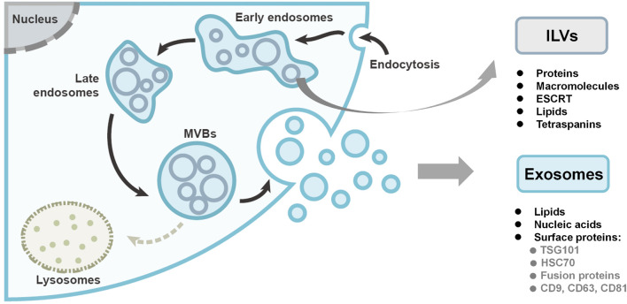
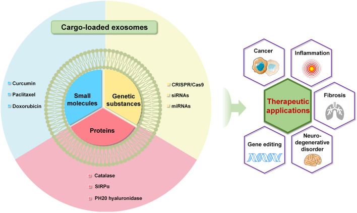
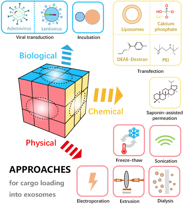
Exosomes are cell-secreted nanoparticles (generally with a size of 30-150 nm) bearing numerous biological molecules including nucleic acids, proteins and lipids, which are thought to play important roles in intercellular communication. As carriers, exosomes hold promise as advanced platforms for targeted drug/gene delivery, owing to their unique properties, such as innate stability, low immunogenicity and excellent tissue/cell penetration capacity. However, their practical applications can be limited due to insufficient targeting ability or low efficacy in some cases. In order to overcome these existing challenges, various approaches have been applied to engineer cell-derived exosomes for a higher selectivity and effectiveness. This review presents the state-of-the-art designs and applications of advanced exosome-based systems for targeted cargo delivery. By discussing experts' opinions, we hope this review will inspire the researchers in this field to develop more practical exosomal delivery systems for clinical applications.

Keywords: drug delivery; exosome engineering; exosomes; extracellular vesicles; gene delivery; targeted delivery.


Conflict of interest statement

The authors declare that the research was conducted in the absence of any commercial or financial relationships that could be construed as a potential conflict of interest.

Figures

FIGURE 1
FIGURE 1
The biogenesis and compositions of exosomes. Exosomes originate as intraluminal vesicles (ILVs) through the inward budding of the plasma membrane within early endosomes. Early endosomes are then matured into late endosomes and ultimately multivesicular bodies (MVBs). Finally, the MVBs either undergo degradation or fuse with the plasma membrane to secrete the ILVs as exosomes. Figure redrawn from Kalluri and LeBleu (2020).
FIGURE 2
FIGURE 2
(A–C) Curcumin encapsulated in exosomes improved the bioavailability after intraperitoneal injection (A); exosomal curcumin reduced lung inflammation (B) and increased survival (C) of lipopolysaccharide-induced septic shock mice. (D,E) Exosome containing SIRPα (Exo-SIRPα) enhanced phagocytosis of HT29 cells by bone marrow derived macrophages (BMDMs) (D) and in vivo antitumor efficacy (E). (F–H) Exosomes loaded with PARP-1 sgR/Cas9 reduced tumor size (F,G) and weight (H) on SKOV3 xenograft mice after intravenous or intratumoral injection (*p < 0.05, **p < 0.01 and ***P < 0.0001). Figure reprinted from Sun et al. (2010); Kim et al. (2017), and Cho et al. (2018).
FIGURE 3
FIGURE 3
Advanced exosome-mediated delivery systems of small molecules, proteins and genetic substances toward various therapeutic applications.
FIGURE 4
FIGURE 4
Schematic diagram of various approaches for cargo loading into or onto exosomes, including physical, chemical, and biological methods.
FIGURE 5
FIGURE 5
Size distribution, zeta potential and morphology of EVs after drug loading with various methods. (A) Size distribution and (B) zeta potential after loading of porBA into vesicles from MDAs. Transmission electron microscopy images of EVs from MDAs indicated that shape and size of EVs after (C) passive, (D) electroporation, and (F) saponin treatment are not altered (arrows). While (E) extrusion, and (G) hypotonic dialysis induced aggregate formation and broader size range of EVs, respectively (arrows) (*p < 0.05). Figure reprinted from Fuhrmann et al. (2015).
FIGURE 6
FIGURE 6
Gene-modified exosomes protect the brain against prolonged deep hypothermic circulatory arrest. (A) Schematic illustration of the principle. (B) Electron microscopic image of miR-214–enriched exosomes. (C) miR-214-3p expression was enhanced significantly in cultured MSCs transfected with premiR-214 (#p < 0.001 vs miR-214-Exo). (D) Intracerebroventricular injection of miR-214–enriched exosomes markedly enhanced the expression of miR-214-3p in the hippocampus (#p < 0.001 vs miR-214-Exo). (E) Number of surviving neurons in CA1 area of hippocampus post administration (*p < 0.001 vs sham group, lp < 0.001 vs control group and Δp < 0.005 vs Exo group or vector-Exo group). Figure reprinted from Shi et al. (2021).
FIGURE 7
FIGURE 7
Modification of exosomes for targeted delivery via direct modification and genetic engineering.

References

    1. Antimisiaris S. G., Mourtas S., Marazioti A. (2018). Exosomes and exosome-inspired vesicles for targeted drug delivery. Pharmaceutics 10:218. 10.3390/pharmaceutics10040218 - DOI - PMC - PubMed
    1. Aqil F., Munagala R., Jeyabalan J., Agrawal A. K., Kyakulaga A. H., Wilcher S. A., et al. (2019). Milk exosomes – natural nanoparticles for siRNA delivery. Cancer Lett. 449 186–195. 10.1016/j.canlet.2019.02.011 - DOI - PubMed
    1. Asadirad A., Hashemi S. M., Baghaei K., Ghanbarian H., Mortaz E., Zali M. R., et al. (2019). Phenotypical and functional evaluation of dendritic cells after exosomal delivery of miRNA-155. Life Sci. 219 152–162. 10.1016/j.lfs.2019.01.005 - DOI - PubMed
    1. Azmi A. S., Bao B., Sarkar F. H. (2013). Exosomes in cancer development, metastasis, and drug resistance: a comprehensive review. Cancer Metastasis Rev. 32 623–642. 10.1007/s10555-013-9441-9 - DOI - PMC - PubMed
    1. Bai J., Duan J., Liu R., Du Y., Luo Q., Cui Y., et al. (2020). Engineered targeting tLyp-1 exosomes as gene therapy vectors for efficient delivery of siRNA into lung cancer cells. Asian J. Pharm. Sci. 15 461–471. 10.1016/j.ajps.2019.04.002 - DOI - PMC - PubMed