Review
. 2022 Mar;40(3):245-261.
doi: 10.1007/s11604-021-01206-5. Epub 2021 Oct 25.

A primer on skeletal dysplasias

Affiliations
Review

A primer on skeletal dysplasias

Atsuhiko Handa et al. Jpn J Radiol. 2022 Mar.

Abstract

Skeletal dysplasia encompasses a heterogeneous group of over 400 genetic disorders. They are individually rare, but collectively rather common with an approximate incidence of 1/5000. Thus, radiologists occasionally encounter skeletal dysplasias in their daily practices, and the topic is commonly brought up in radiology board examinations across the world. However, many radiologists and trainees struggle with this issue because of the lack of proper resources. The radiological diagnosis of skeletal dysplasias primarily rests on pattern recognition-a method that is often called the "Aunt Minnie" approach. Most skeletal dysplasias have an identifiable pattern of skeletal changes composed of unique findings and even pathognomonic findings. Thus, skeletal dysplasias are the best example to which the Aunt Minnie approach is readily applicable.

Keywords: Aunt Minnie; Radiology board examination; Skeletal dysplasia.


Conflict of interest statement

The authors declare that there is no conflict of interest.

Figures
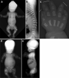
Fig. 1
Fig. 1
a, b A neonate with achondroplasia. a Radiographs show a disproportionately large skull, a narrow thorax, absence of normal increase in caudal interpedicle distances, small iliac bones with short sacrosciatic notches (asterisks) and trident-appearing acetabula (corniculate protrusion of the lateral, middle, and medial edge of the triradiate cartilage; arrows), ovoid lucency in the proximal femora, short and broad tubular bones associated with metaphyseal cupping, lateral slanting of the end of the distal femoral ends (arrowhead), short tibiae leading to relative elongation of the fibulae, and b “bullet-shaped” flat vertebral bodies and narrow spinal canal (on lateral view). c A frontal hand radiograph of another neonate with achondroplasia shows radial slanting of the 2nd metacarpal end and proximal phalangeal base, and a so-called “trident hand” (unable to oppose the 3rd and 4th fingers; three white lines). d, e A stillbirth with thanatophoric dysplasia. Radiographs show a disproportionately large skull, an extremely narrow thorax, severe platyspondyly (i.e., flat spine), caudally decreased interpedicle distances, small iliac bones with narrow sacrosciatic notches and trident-shaped acetabula, bowed femora (termed “French telephone receiver femora”), ovoid lucency of the proximal humeri/ femora (caused by metaphyseal cupping), metaphyseal cupping of the long bones. Note all the skeletal changes are qualitatively similar to, but quantitatively much more severe than those of achondroplasia
Fig. 2
Fig. 2
An infant with chondrodysplasia punctata. Multiple puncta are seen in the tarsal bones and the phalanges
Fig. 3
Fig. 3
a A neonate with Jeune asphyxiating thoracic dysplasia. Radiograph shows markedly short ribs, and trident-shaped ilia (arrows). Severe brachydactyly without polydactyly were also noted (not shown). Premature ossification of the proximal femoral epiphyses is distinctive. As opposed to achondroplasia, there is no caudally decreased interpedicle distance or ovoid lucency of the proximal femora. (Reprinted from Handa, A, et al. Jpn J Radiol. 2020;38(3):193–206). b A child with the same diagnosis. Radiograph shows cone-shaped epiphyses involving both the phalanges and the metacarpals. Cone-shaped phalangeal epiphyses and inverted tear-shaped metacarpal epiphyses invaginate into cupped metaphyses. Premature fusion of the growth plate is also seen in the middle and distal phalanges. (Reprinted from Handa, A, et al. Jpn J Radiol. 2020;38(3):193–206)
Fig. 4
Fig. 4
A 5-year-old child with multiple epiphyseal dysplasia, autosomal dominant type. Radiographs show delayed, disorganized epiphyseal ossification including small and round capital femoral epiphyses and irregular epiphyseal ossification of the knee
Fig. 5
Fig. 5
ac A neonate with SEDC. Radiographs show a broad thorax and modest platyspondyly (flattening of the vertebral bodies) due to delayed vertebral ossification. The delayed ossification is prominent in the dorsal vertebral bodies, giving rise to pear-shaped vertebral bodies seen on the lateral view. The delayed ossification is more severe in the cervical spine and sacrum than in the thoracolumbar spine, creating anisospondyly (increased variability of the size of the vertebral bodies). The ilia are craniocaudally short, the pubic bones are absent, epiphyseal ossification of the distal femora is retarded (typically seen by 39 weeks of gestation), and the long bones show mild metaphyseal widening
Fig. 6
Fig. 6
ad An 11-year-old boy with Hurler syndrome. Radiographs show findings of dysostosis multiplex including a thick clavicles, paddle-shaped (or oar-shaped) broad ribs, b comma-shaped ilia, diaphyseal broadening and metaphyseal constriction of the long bones, c hook-shaped vertebral bodies (arrows) with posterior scalloping, d proximal pointing of the metacarpals (arrowhead), and bullet-shaped phalanges
Fig. 7
Fig. 7
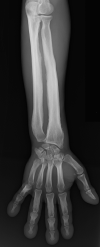
a, b A 12-year-old girl with Albright hereditary osteodystrophy. Radiographs show shortening of the ulnar metacarpals (type E brachydactyly) and short distal phalanges, particularly of the thumb, and periarticular soft tissue calcifications around both knees (arrows)
Fig. 8
Fig. 8
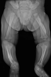
a A stillborn with osteogenesis imperfecta. Radiograph shows a beaded appearance of the ribs and an “accordion-like” wavy appearance of the long bones as a consequence of in utero multiple fractures. b, c A 2-year-old patient with osteogenesis imperfecta. Radiographs show multiple Wormian bones (arrow) due to defective calvarial ossification and generalized osteoporosis with a healing fracture of the right femoral shaft and a healed fracture of the left femoral shaft
Fig. 9
Fig. 9
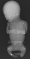
A stillborn with hypophosphatasia. Radiograph shows near-absent ossification of the skull (yet the frontal bones are preserved), short and thin ribs, small scapulae, absent ossification of the pubic and ischial bones, and sharp angulation of the long bones with metaphyseal defects extending into the diaphysis. Vertebral ossification is unequal. Some vertebral bodies are completely unossified, while others are well-ossified. The neural arches are thoroughly missing
Fig. 10
Fig. 10
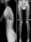
a, b A 14-year-old boy with adult type osteopetrosis. Radiographs show generalized increased bone density with poor corticomedullary differentiation and mild Erlenmeyer flask deformity of the long bones (arrows). Lateral view of the spine shows prominent superior and inferior endplate sclerosis, giving rise to a sandwich appearance of the vertebral bodies
Fig. 11
Fig. 11
ac A young adult with pyknodysostosis. Radiographs show craniofacial sclerosis with widely opened fontanels, severe micrognathia with an obtuse mandibular angle, and edentulous state due to repeated dental infections as well as generalized increased bone density with a preserved corticomedullary differentiation and trabecular pattern, healed fractures of both tibiae and c severe acroosteolysis (arrowheads)
Fig. 12
Fig. 12
a A 10-year-old boy with osteopoikilosis. Radiograph shows small foci of bone sclerosis in different sizes and shapes (round, oval, lenticular) located in the pelvis and bilateral proximal femora. b A 4-year-old girl with osteopathia striata. Radiograph shows longitudinal striations in the distal femoral and proximal tibial metaphyses. c A 75-year-old male with melorheostosis. Radiograph shows dripping candle wax-like, eccentric hyperostoses distributed along the tibia
Fig. 13
Fig. 13
a, b An adult patient with metaphyseal dysplasia (Pyle disease). Radiographs show broad clavicles and ribs as well as significant expansion and thin cortices of the metaphysis and metadiaphyseal junction of the long bones giving rise to “Erlenmeyer flask” deformities. There are fractures of the right distal tibia and fibula
Fig. 14
Fig. 14
a, b A 3-year-old girl with diaphyseal dysplasia (Camurati–Engelmann disease). Radiograph shows cortical thickening with coinciding medullary narrowing of the long bones
Fig. 15
Fig. 15
A 26-year-old male with pachydermoperiostosis. Radiograph shows irregular periosteal reaction of the short tubular bones and the long bones. Digital clubbing is also seen
Fig. 16
Fig. 16
A 2-month-old infant with infantile cortical hyperostosis (Caffey disease). Radiograph shows cortical hyperostosis in the left femur and right tibia
Fig. 17
Fig. 17
a, b A 25-year-old female with cleidocranial dysplasia. Radiographs show multiple Wormian bones, absent bilateral clavicles, and a narrow upper thorax
Fig. 18
Fig. 18
a, b A 28-year-old female with Nail–Patella syndrome. There are pathognomonic iliac horns (arrows) and hypoplastic patellae
Fig. 19
Fig. 19
a A child with hereditary multiple exostoses. There are multiple exostoses (pedunculated bony protrusions with corticomedullary continuation) of the metaphyses of the long bones. b A 20-year-old female with hereditary multiple exostoses. There are sessile exostoses with corticomedullary continuation of the right humerus which impaired the growth of the proximal humeral growth plate
Fig. 20
Fig. 20
A 5-year-old boy with dysplasia epiphysealis hemimelica (Trevor’s disease). Radiograph shows osseous overgrowth arising from the right medial femoral condyle
Fig. 21
Fig. 21
a An 8-year-old child with enchondromatosis (Ollier disease). Radiograph shows multiple expansile metaphyseal lucent lesions in the third to fifth phalanges. b A young adult with Maffucci syndrome. Radiograph shows multiple enchondromatosis in the second to fourth digits with soft tissue masses associated with multiple foci of round phleboliths compatible with hemangiomas
Fig. 22
Fig. 22
a A 13-year-old boy with polyostotic fibrous dysplasia accompanied by café-au-lait spots. Radiograph shows elongated and expansile “ground glass” lesions replacing the bone marrow in the proximal femoral diaphysis and the tibial diaphysis. b, c A 59-year-old male with Mazabraud syndrome. Radiograph shows fibrous dysplasia involving the femur with coxa varus angulation, giving what is called a “shepherd crook deformity”. Coronal STIR image showing intramuscular hyperintensity compatible with intramuscular myxomas (arrows) as well as heterogeneous hyperintense signal in the femur compatible with fibrous dysplasia (arrowhead)

References

    1. Langer LO, Jr, Baumann PA, Gorlin RJ. Achondroplasia. Am J Roentgenol Radium Ther Nucl Med. 1967;100(1):12–26. - PubMed
    1. Bellus GA, Hefferon TW, Ortiz de Luna RI, Hecht JT, Horton WA, Machado M, et al. Achondroplasia is defined by recurrent G380R mutations of FGFR3. Am J Hum Genet. 1995;56(2):368–373. - PMC - PubMed
    1. Tavormina PL, Shiang R, Thompson LM, Zhu YZ, Wilkin DJ, Lachman RS, et al. Thanatophoric dysplasia (types I and II) caused by distinct mutations in fibroblast growth factor receptor 3. Nat Genet. 1995;9(3):321–328. - PubMed
    1. Maroteaux P, Lamy M, Robert JM. Thanatophoric dwarfism. Presse Med. 1967;75(49):2519–2524. - PubMed
    1. Spranger JW, Opitz JM, Bidder U. Heterogeneity of Chondrodysplasia punctata. Humangenetik. 1971;11(3):190–212. - PubMed

LinkOut - more resources