. 2022 Jan;91(1):101-116.
doi: 10.1002/ana.26256. Epub 2021 Nov 16.

Cardiac Investigations in Sudden Unexpected Death in DEPDC5-Related Epilepsy

Collaborators, Affiliations

Cardiac Investigations in Sudden Unexpected Death in DEPDC5-Related Epilepsy

Alexandre Bacq et al. Ann Neurol. 2022 Jan.

Abstract

Objective: Germline loss-of-function mutations in DEPDC5, and in its binding partners (NPRL2/3) of the mammalian target of rapamycin (mTOR) repressor GATOR1 complex, cause focal epilepsies and increase the risk of sudden unexpected death in epilepsy (SUDEP). Here, we asked whether DEPDC5 haploinsufficiency predisposes to primary cardiac defects that could contribute to SUDEP and therefore impact the clinical management of patients at high risk of SUDEP.

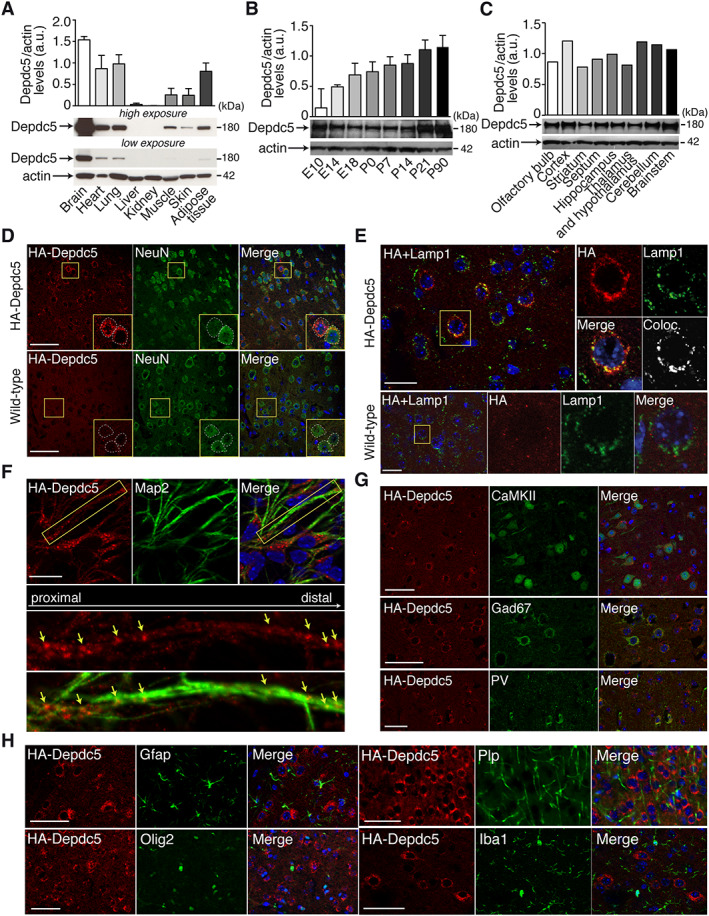
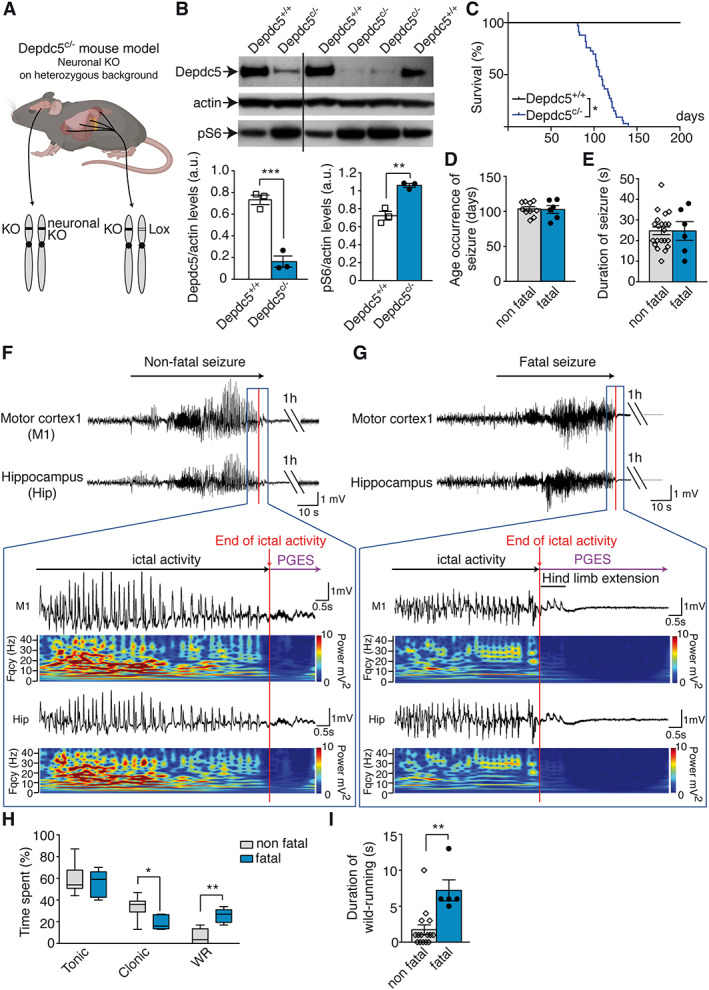
Methods: Clinical cardiac investigations were performed in 16 patients with pathogenic variants in DEPDC5, NPRL2, or NPRL3. Two novel Depdc5 mouse strains, a human HA-tagged Depdc5 strain and a Depdc5 heterozygous knockout with a neuron-specific deletion of the second allele (Depdc5c/- ), were generated to investigate the role of Depdc5 in SUDEP and cardiac activity during seizures.

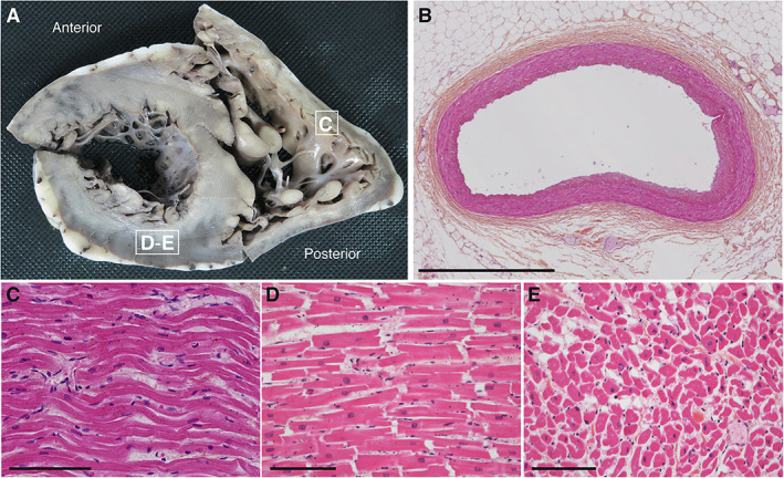
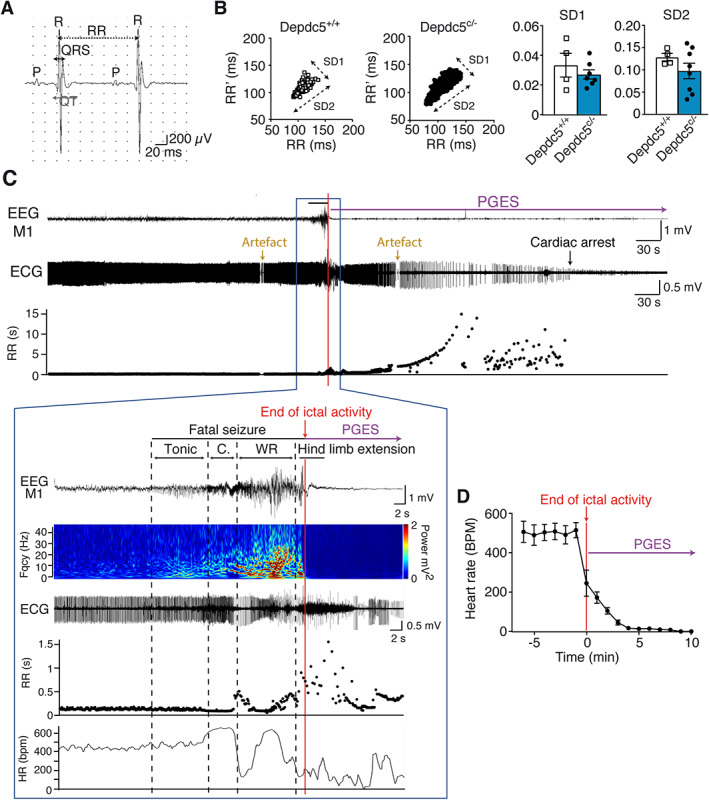
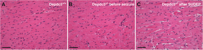
Results: Holter, echocardiographic, and electrocardiographic (ECG) examinations provided no evidence for altered clinical cardiac function in the patient cohort, of whom 3 DEPDC5 patients succumbed to SUDEP and 6 had a family history of SUDEP. There was no cardiac injury at autopsy in a postmortem DEPDC5 SUDEP case. The HA-tagged Depdc5 mouse revealed expression of Depdc5 in the brain, heart, and lungs. Simultaneous electroencephalographic-ECG records on Depdc5c/- mice showed that spontaneous epileptic seizures resulting in a SUDEP-like event are not preceded by cardiac arrhythmia.

Interpretation: Mouse and human data show neither structural nor functional cardiac damage that might underlie a primary contribution to SUDEP in the spectrum of DEPDC5-related epilepsies. ANN NEUROL 2022;91:101-116.


Conflict of interest statement

Nothing to report.

Figures

FIGURE 1
FIGURE 1
Macroscopic and microscopic pictures of the heart autopsy of Patient 1. (A) Short axis view of a transverse section of the right and left ventricles. Morphological cardiovascular examination including valves and coronary arteries inspection revealed no gross abnormalities. White squares indicate sites of sections used for histological analysis in C–E. (B–E) Hematoxylin and eosin–stained transversal sections of the anterior interventricular artery (B), longitudinal sections of the right ventricle myocardium (C), the posterior wall of the left ventricle myocardium (D), and cross‐sectional section of the left ventricle (E). No contraction band necrosis was detected. Scale bars represent 1mm (B) or 100μm (C–E).
FIGURE 2
FIGURE 2
Depdc5 expression in the HA‐tagged Depdc5 mouse. (A–C) Quantified representative Western blots show HA‐Depdc5 expression in (A) various mouse organ lysates, (B) mouse brain lysates at developmental stages from embryonic day 10 (E10) to postnatal day 90 (P90), and (C) different brain regions of adult mouse (n = 1–3). Actin was used as the loading control. (D–H) Depdc5 expression in the somatosensory cortex. (D) Immunofluorescent colabeling of HA‐Depdc5 (red) with NeuN (green) showing specific expression of Depdc5 (compared to wild‐type untagged cortex, bottom) in neuronal soma (see insets, corresponding to the yellow squares). (E) Immunofluorescent colabeling of HA‐Depdc5 (red) with Lamp1 (green) showing specific enriched expression in lysosomes (compared to wild‐type untagged cortex, bottom). On the right of insets (corresponding to the yellow square) are the colocalization (Coloc.) between HA and Lamp1 (Pearson correlation coefficient for HA‐Depdc5 mouse = 0.49). (F) Immunofluorescent colabeling of HA‐Depdc5 (red) with Map2 (green) showing expression of Depdc5 in neurites. Bottom images are the insets (corresponding to the yellow square) showing HA (red) distribution along the MAP2‐positive (green) neurite. Arrows show aggregation of HA‐Depdc5 staining. (G) Immunofluorescent colabeling of HA‐Depdc5 (red) and CaMKII (excitatory neurons), Gad67 (inhibitory neurons) or parvalbumin (PV; PV interneurons) shows Depdc5 is expressed in excitatory and inhibitory neurons. (H) Colabeling of HA‐Depdc5 (red) and Gfap, Olig2, Plp, and Iba1, shows no coexpression in glial and microglial cells. Three sections were done in duplicate. Scale bars represent 50μm (D, G, H), 25μm (E), or 20μm (F).
FIGURE 3
FIGURE 3
Spontaneous seizures in Depdc5 c/− mice. (A) Illustration of the Depdc5 c/− mouse model generated. KO = knockout. (B) Representative Western blot of brain lysates from Depdc5 c/− and control wild type (Depdc5 +/+) immunostained for Depdc5, actin, and pS6 (top) and quantification (bottom) of Depdc5 levels (left, unpaired t test, t 4 = 8.4, p = 0.0006) and pS6 levels (right, unpaired t test, t 4 = 6.4, p = 0.0031). (C) Survival of Depdc5 c/− mice (n = 33, log‐rank test, p = 0.047). (D) Age at onset of nonfatal (n = 6) and fatal seizures (n = 10, unpaired t test, t 14 = 0.17, p = 0.87). (E) Duration of nonfatal (n = 6) and fatal seizures (n = 21, unpaired t test, t 25 = 0.011, p = 0.99). (F) Representative electroencephalographic (EEG) recordings and fast Fourier transform (FFT) power spectrum of nonfatal seizure in a Depdc5 c/− mouse, followed with a postictal generalized EEG suppression (PGES) of 3 minutes 34 seconds. The color‐coded FFT power spectrum shows EEG amplitude and frequency changes from motor cortex (M1) and hippocampus (Hip). (G) Representative EEG recordings and FFT of a fatal seizure in a Depdc5 c/− mouse terminating with hind limb extension and prolonged PGES. (H) Duration of tonic, clonic, and wild running (WR) phases during nonfatal and fatal seizures in Depdc5 c/− mice (n = 5–12; 2‐way analysis of variance revealed main effect of behavior type, F 2,45 = 62.85, p < 0.0003; and main effect of interaction, F 2,45 = 9.98, p = 0.0003; Bonferroni post hoc tests). (I) Duration of wild running in nonfatal and fatal seizures (n = 5–21, unpaired t test, t 24 = 3.83, p = 0.0012). Results are given as mean ± standard error of the mean. *p < 0.05, **p < 0.01, ***p < 0.001 versus Depdc5 +/+.
FIGURE 4
FIGURE 4
Electroencephalographic (EEG) and electrocardiographic (ECG) records from Depdc5 c/− mice at rest and during a spontaneous fatal seizure. (A) Example of ECG records in anesthetized mice. (B) In vivo recordings of heart rate variability are represented with Poincaré plots in Depdc5 c/− and Depdc5 +/+ mice, and quantified by SD1 (Mann–Whitney, U = 7, p = 0.53) and SD2 (Mann–Whitney, U = 11, p = 0.46) parameters (n = 4–8 mice, n > 500 RR measures per animal). (C) Representative simultaneous EEG‐ECG records and RR plot before, during, and after a fatal seizure in a Depdc5 c/− mouse, terminating with prolonged postictal generalized EEG suppression (PGES). Fast Fourier transform power spectrum shows EEG amplitude and frequency changes from motor cortex (M1). Dashed lines indicate the different behavioral phases (Tonic, C = Clonic, and WR = Wild Running). RR length and heart rate (HR; mean number of RR in 2 seconds) changed after the beginning of the seizure, during the clonic phase. The ECG during the seizure and after the hind limb extension phase is partially obscured by electrical activity of muscle contraction. Artifacted RR measures were excluded. (D) Average heart rate changes during and after the seizure in 6 Depdc5 c/− mice. Results are mean ± standard error of the mean.
FIGURE 5
FIGURE 5
Mouse heart histology before and after sudden unexpected death in epilepsy (SUDEP)‐like event. Hematoxylin and eosin–stained transversal sections of the anterior interventricular artery are shown in a control Depdc5 +/+ (A), Depdc5 c/− before seizure (B), and Depdc5 c/− after SUDEP‐like event (C). No contraction band necrosis or fibrosis was detected in Depdc5 c/− heart after SUDEP. Scale bars represent 100μm.

References

    1. Nashef L, So EL, Ryvlin P, Tomson T. Unifying the definitions of sudden unexpected death in epilepsy. Epilepsia 2012;53:227–233. - PubMed
    1. Harden C, Tomson T, Gloss D, et al. Practice guideline summary: sudden unexpected death in epilepsy incidence rates and risk factors: report of the Guideline Development, Dissemination, and Implementation Subcommittee of the American Academy of Neurology and the American Epilepsy Society. Epilepsy Curr 2017;17:180–187. - PMC - PubMed
    1. Whitney R, Donner EJ. Risk factors for sudden unexpected death in epilepsy (SUDEP) and their mitigation. Curr Treat Options Neurol 2019;21:7. - PubMed
    1. Ryvlin P, Nashef L, Lhatoo SD, et al. Incidence and mechanisms of cardiorespiratory arrests in epilepsy monitoring units (MORTEMUS): a retrospective study. Lancet Neurol 2013;12:966–977. - PubMed
    1. Devinsky O, Hesdorffer DC, Thurman DJ, et al. Sudden unexpected death in epilepsy: epidemiology, mechanisms, and prevention. Lancet Neurol 2016;15:1075–1088. - PubMed

Publication types